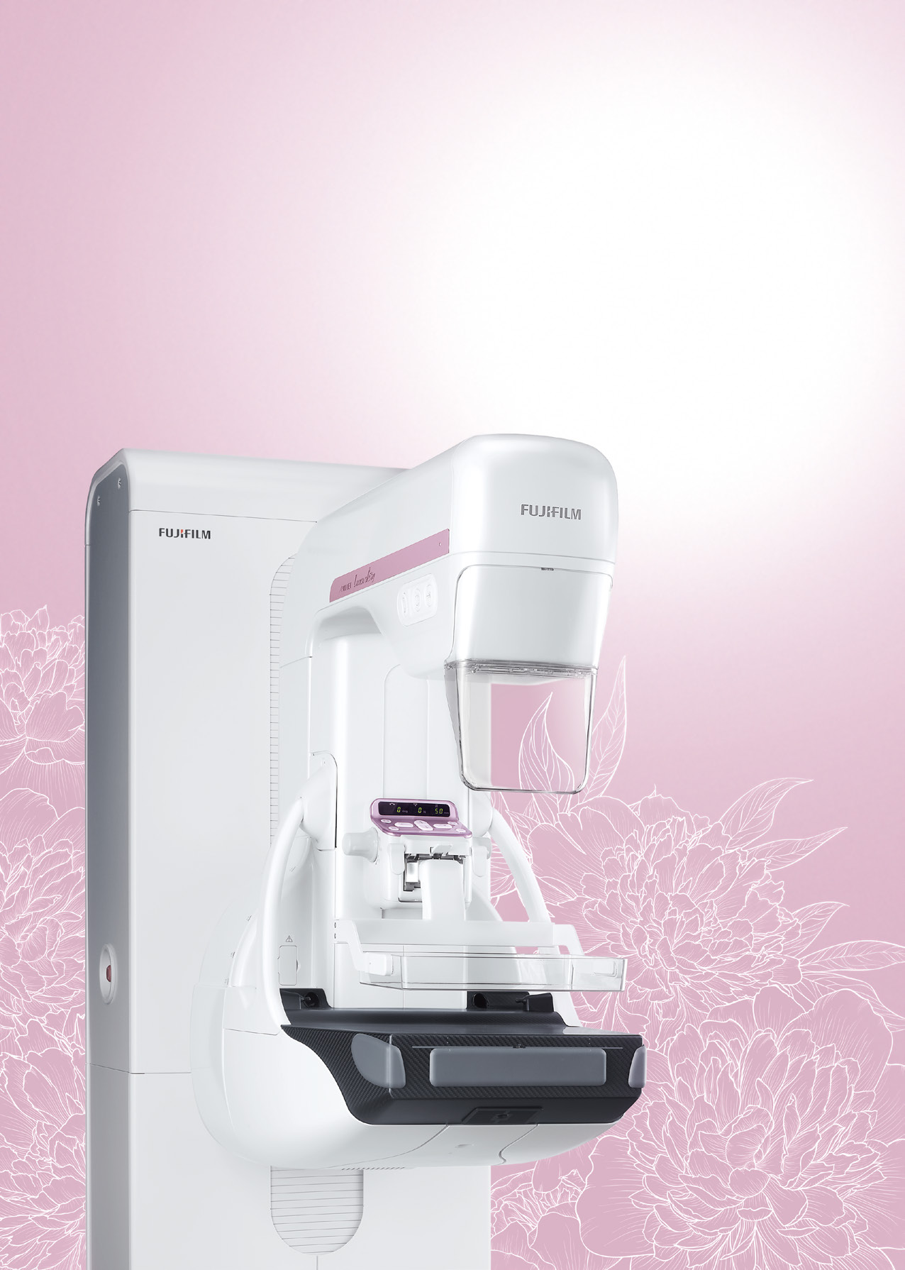
October is Breast Cancer Awareness Month. If you are 40 or over, ask your doctor whether it is time for your mammogram.
Digital breast tomosynthesis (3D mammography) using the Fujifilm Amulet Innovality — detects up to 40% more cancers than standard 2D mammography. BI-RADS structured reporting by our consultant radiologist for breast and women's imaging.
Fujifilm Amulet InnovalityBI-RADS Reporting3D TomosynthesisBreast Sonography IncludedMost Reports Same Day
AV Scan Diagnostics in Dadar West / Prabhadevi, Mumbai, near Siddhivinayak Temple and Shivaji Park, provides 3D mammography (digital breast tomosynthesis) on a Fujifilm Amulet Innovality. Every mammogram here is paired with a breast ultrasound performed by our breast imaging specialist, MD (Radiology). She is a woman doctor and does every breast ultrasound here personally, including evenings and Sundays. Reports use the BI-RADS system and most are ready the same day, sent to your WhatsApp; if a finding needs a sample, a same-day ultrasound-guided biopsy is possible.
At a Glance
What it is
3D mammography (digital breast tomosynthesis): a low-dose breast X-ray taken from several angles and rebuilt into thin slices
Equipment
Fujifilm Amulet Innovality 3D mammography (tomosynthesis)
Paired with
A breast ultrasound (sonomammography) with every mammogram, in line with BI-RADS practice, performed by our breast imaging specialist herself
Reported by
Our breast imaging specialist, MD (Radiology), FMF (UK) certified
Typical time
The mammogram usually takes about 15-20 minutes; the breast ultrasound usually about 15-30 minutes more
Preparation
No fasting; no deodorant, antiperspirant, talc or powder on the day; wear a two-piece outfit; bring previous mammograms; tell us about implants, pregnancy or breastfeeding. Our team confirms the exact preparation when you book on WhatsApp
Report
BI-RADS report; most reports the same day, sent to your WhatsApp; images by cloud link or DVD
Price
The exact price is confirmed on WhatsApp before your visit
Centre hours
Mon-Sat 8am-10pm, Sun 8am-5pm
Location and access
Ground Floor, Shilpa Apartment, Shankar Ghanekar Marg, Dadar West / Prabhadevi, Mumbai 400028, near Siddhivinayak Temple and Shivaji Park; step-free
3D Mammography at AV Scan Diagnostics, Dadar West
AV Scan Diagnostics offers 3D digital breast tomosynthesis using the Fujifilm Amulet Innovality. Unlike standard 2D mammography, tomosynthesis takes a series of low-dose images of the breast from different angles and rebuilds them into thin slices, so our radiologist can examine the breast tissue layer by layer.
All mammography reports at AV Scan are personally reviewed and signed by our consultant radiologist for breast and women's imaging — MD (Radiology), FIFM, FMF (UK) certified — using BI-RADS (Breast Imaging-Reporting and Data System) standardised reporting.
In large studies, adding tomosynthesis to mammography found more cancers and led to fewer call-backs for extra views than 2D mammography alone. The gain is seen in many women with dense breasts but is smaller in extremely dense breasts, which is one reason every mammogram at AV Scan is also paired with a breast ultrasound.
Who Should Get a Mammogram?
Women aged 40 and above, for routine screening, usually every one to two years; your doctor advises the interval that suits you
Women with a family history of breast cancer
Women with dense breast tissue, where 3D mammography and the paired breast ultrasound both help
Women with BRCA1/BRCA2 gene mutations
Anyone with a breast lump, nipple discharge, or breast skin changes
Women undergoing breast implant assessment
Women referred for baseline mammography before hormone therapy
3D vs 2D Mammography — What's the Difference?
Standard 2D mammography takes flat images of the breast from two angles. Overlapping tissue can mask cancers or create suspicious areas that turn out to be normal tissue.
3D mammography (tomosynthesis) acquires multiple low-dose images from different angles and reconstructs them into thin slices, allowing the radiologist to scroll through the breast tissue layer by layer. This reduces the effect of overlapping tissue; in large studies it found more cancers and led to fewer call-backs than 2D mammography alone.
2D mammography
3D mammography (tomosynthesis)
How the pictures are taken
Two flat images of each breast
Many low-dose images from different angles, rebuilt into thin slices
Overlapping breast tissue
Can hide a small cancer, or look like one
The radiologist scrolls through the breast layer by layer, so overlap matters less
Call-backs for extra views
More frequent
Fewer in large studies
Dense breasts
Harder to read
Often easier to read; at AV Scan every mammogram is also paired with breast ultrasound
What you feel
Breast compression for a few seconds per view
The same compression, a few seconds longer per view
Radiation
Low dose
Low dose, in a similar range (depends on the technique used)
Breast Sonography (Sonomammography) — Always Included
At AV Scan, every mammography examination is routinely followed up with breast sonography (sonomammography) — a complementary ultrasound of the breast performed immediately after your mammogram. This is our standard protocol, in line with BI-RADS practice, not an optional add-on.
Breast ultrasound is especially valuable for:
Women with dense breast tissue, where ultrasound can find some cancers that the mammogram does not show
Evaluating any area that looks suspicious on the mammogram
Distinguishing solid lumps from fluid-filled cysts
Guiding further assessment or biopsy if needed
Younger women where ultrasound is often the primary imaging tool
Our combined 3D mammography + breast sonography protocol gives you a more complete breast assessment in one visit. The ultrasound can find some cancers that are hidden in dense tissue; it also shows more harmless spots, and your report states clearly which findings, if any, need a follow-up scan.
Dense Breast Screening — What Your Report Means
Breast density describes how much of the breast is gland and fibrous tissue rather than fat. It is common, especially before menopause, and it is not a disease. Following BI-RADS, a mammogram report places density in one of four groups, from A (almost entirely fatty) to D (extremely dense). Groups C and D are called dense breasts.
Density matters for two reasons. Dense tissue and a cancer both look white on a mammogram, so a small cancer can be harder to see. Dense breasts are also linked with a somewhat higher risk of breast cancer. At AV Scan Diagnostics in Dadar West, Mumbai, dense breasts are checked in complementary ways:
3D tomosynthesis, so the radiologist can look through overlapping tissue layer by layer. This helps in many women with dense breasts, though the gain is smaller when the breasts are extremely dense.
A breast ultrasound with every mammogram, performed by our breast imaging specialist herself. It can find some cancers hidden in dense tissue, and it also shows more harmless spots that may need a follow-up scan. Read more about breast ultrasound (sonomammography).
Breast MRI when your doctor advises it, for example for women at high risk, some women with extremely dense breasts, or when mammography and ultrasound leave a question open. Read more about breast MRI.
No single test finds every cancer. If your report says you have dense breasts, discuss with your doctor whether extra screening suits your personal and family risk. Bring your earlier mammograms, CDs and reports, because comparing with previous years makes dense breasts easier to read.
How to Prepare for Your Mammogram
If you still have periods, the week after your period is often more comfortable, as the breasts are less tender. Do not delay a scan for a new lump.
Do not apply deodorant, antiperspirant, perfume or powder on the day of your scan — these can interfere with the images.
Wear a two-piece outfit for easy access.
Bring any previous mammogram images for comparison.
Tell us about any breast implants, previous breast surgery or biopsy.
Tell us if you are, or might be, pregnant, or if you are breastfeeding.
Our team confirms the exact preparation when you book on WhatsApp.
What to Expect During Your 3D Mammogram
Your 3D mammogram at AV Scan Diagnostics is taken on the Fujifilm Amulet Innovality and is followed by a breast ultrasound in the same visit. A typical visit goes like this:
Before the scan: our team asks about any symptoms, earlier breast surgery, biopsy or implants, and whether you are or might be pregnant, and collects your previous mammograms and reports for comparison.
Changing: you undress from the waist up and wear a gown.
The mammogram: each breast in turn is placed on the detector and pressed gently but firmly by a clear plastic paddle, usually for two views of each breast, one from above and one from an angle at the side. For each 3D view, the X-ray tube moves in a short arc over the breast for a few seconds while you hold still. Sometimes extra views are needed, for example if you have breast implants.
Compression: many women feel firm pressure or discomfort for a few seconds per view rather than lasting pain. Compression spreads the tissue so the pictures are clear and the dose stays low. Tell the radiographer at once if it hurts.
The breast ultrasound: our breast imaging specialist then scans both breasts and armpits herself, looking closely at any area that worries you or that the mammogram shows.
Time: the mammogram usually takes about 15-20 minutes and the ultrasound usually about 15-30 minutes more, plus time to change.
Afterwards: you can usually go back to your normal day straight away. If a biopsy or FNAC is done in the same visit, our team gives you separate aftercare advice. Your report uses BI-RADS categories. Most reports are ready the same day and sent to your WhatsApp, and images are shared by cloud link or DVD.
How to Choose Where to Have Your Mammogram in Mumbai
Instead of relying on rankings, check a few neutral points before you book, wherever you go:
Whether 3D mammography (tomosynthesis) is available, particularly if you have been told you have dense breasts.
Who reports the mammogram, and whether their work focuses on breast imaging.
Whether the report uses BI-RADS categories and states your breast density.
Whether a breast ultrasound can be done in the same visit, and who performs it.
Whether a biopsy can be arranged promptly if something needs a sample.
Whether your previous mammograms will be compared with the new one.
How and when you receive your report and images.
At AV Scan Diagnostics in Dadar West / Prabhadevi, Mumbai, 3D mammography is done on the Fujifilm Amulet Innovality. Every mammogram is paired with a breast ultrasound performed by our breast imaging specialist, MD (Radiology), FMF (UK) certified. Reports use BI-RADS, and most are ready the same day on WhatsApp. A same-day ultrasound-guided biopsy or FNAC is possible when needed. The centre was co-founded by two MD radiologists. It is open 7 days a week, on the ground floor with step-free access, near Siddhivinayak Temple and Shivaji Park. It is rated 4.9★ on Google.
Breast FNAC and Biopsy — Done Here
If your mammogram or sonography shows something that needs a tissue diagnosis, it can be done in the same building rather than sending you somewhere else. We perform ultrasound-guided breast FNAC (fine needle aspiration cytology) and breast biopsy on site. The procedure is performed by our radiologists; the sample is then examined by a pathologist at our partner laboratory, who issues the result.
Breast FNAC — a fine needle sample taken under ultrasound guidance, with a local anaesthetic where needed. It usually takes a few minutes.
Breast biopsy — a core tissue sample, for when cytology alone is not enough.
Axillary lymph node FNAC — for nodes picked up on imaging.
When needed, the FNAC or biopsy can be done on the same day as your mammogram and sonography.
Message us on WhatsApp and we will tell you what applies and what it costs before you travel.
Frequently Asked Questions — Mammography in Mumbai
Many women feel firm pressure or some discomfort rather than pain, and it lasts a few seconds per view; a few find it painful, especially when the breasts are tender. Compression spreads the tissue so the pictures are clear and the radiation dose stays low. You can tell the radiographer at any time if it hurts, and if you still have periods, booking for the week after your period often helps.
3D mammography uses a low dose of X-rays. Depending on how the views are taken, the dose is similar to or somewhat higher than a standard 2D mammogram, and it stays within the limits set for mammography. For women of screening age, the benefit of finding a cancer early is generally considered to outweigh the very small risk from this dose. If you are, or might be, pregnant, tell us before the scan.
BI-RADS (Breast Imaging-Reporting and Data System) is a widely used standard system for breast imaging reports, developed by the American College of Radiology. It assigns categories from 0–6 indicating the likelihood of malignancy and recommended next steps. All mammography reports at AV Scan are BI-RADS compliant and signed by our breast imaging specialist.
Often, yes. Dense tissue and cancers both look white on a mammogram, so overlapping tissue can hide a small cancer on 2D images. Tomosynthesis lets the radiologist look through the breast layer by layer, which helps in many women with dense breasts, though the gain is smaller in extremely dense breasts. That is one reason every mammogram at AV Scan is also paired with a breast ultrasound by our breast imaging specialist. Discuss with your doctor whether breast MRI is also advisable for your level of risk.
AV Scan Diagnostics does not publish prices online, because the cost depends on what your doctor has asked for: a routine screening or a check of a specific problem, whether extra views are needed, and whether a biopsy is needed on the day. Every mammogram here is paired with a breast ultrasound by our breast imaging specialist. Send your prescription on WhatsApp to +91 98200 51511: the exact price is confirmed on WhatsApp before your visit. See how our prices are quoted. For insurance reimbursement, we give you every bill and report your claim needs.
Many guidelines suggest routine screening mammograms from about age 40, usually every one to two years. Guidelines differ, and the right age and interval depend on your health and family history, so discuss it with your doctor. Women at higher risk, such as those with a BRCA1 or BRCA2 gene change or a strong family history, may be advised to start earlier, sometimes with yearly breast MRI as well. In younger women, a breast lump is usually checked with ultrasound first. October is Breast Cancer Awareness Month and a good reminder to ask, but a new lump, nipple discharge or skin change should be checked promptly in any month, whatever your age and even if your last mammogram was normal.
Many findings turn out to be harmless. Every mammogram at AV Scan Diagnostics is paired with a breast ultrasound by our breast imaging specialist, so the ultrasound often answers the question in the same visit. The report gives a BI-RADS category: category 0 means more pictures are needed before a final answer, category 3 (probably benign) usually means a follow-up scan in about 6 months, and category 4 or 5 means a biopsy is advised. An ultrasound-guided biopsy or FNAC is possible the same day when needed, and a pathologist at our partner laboratory issues the result. Your doctor then explains the next step.
They see different things, so neither replaces the other. A mammogram shows tiny specks of calcium (microcalcifications), which can be an early sign of some breast cancers, and ultrasound does not reliably show them. Ultrasound can tell a fluid-filled cyst from a solid lump and can find some cancers hidden in dense tissue. For routine screening, usually from about 40, the mammogram is the main test; in younger women with a lump, ultrasound usually comes first. At AV Scan Diagnostics, every mammogram is paired with a breast ultrasound. See breast ultrasound (sonomammography).
Tell us before booking if you are, or might be, pregnant, or if you are breastfeeding. For a breast lump during pregnancy or breastfeeding, breast ultrasound is usually the first test, and our breast imaging specialist performs it herself. A mammogram can still be done if the ultrasound shows something that needs it; the X-rays are aimed at the breast and the dose reaching the baby is very small. If you are breastfeeding, feeding or expressing shortly before the scan makes it more comfortable and the pictures clearer. A routine screening mammogram is usually timed for after pregnancy, so discuss the timing with your doctor.
AV Scan Diagnostics is at Ground Floor, Shilpa Apartment, Shankar Ghanekar Marg, Dadar West / Prabhadevi, Mumbai 400028, near Siddhivinayak Temple and Shivaji Park. The centre is on the ground floor with step-free access and is open Monday to Saturday 8am-10pm and Sunday 8am-5pm. Book your mammogram slot on WhatsApp at +91 98200 51511 or call 022-3522 0711. Directions are on our Visit Us page.