At a Glance
| Also called | Ankle MRI, foot MRI, MRI heel; prescriptions usually name the side, such as "MRI right ankle" |
|---|
| Radiation and contrast | No radiation; contrast usually not needed |
|---|
| Preparation | Usually none; eat and take your medicines as normal (confirmed on WhatsApp) |
|---|
| Scan time | Usually about 20-40 minutes for one ankle or foot |
|---|
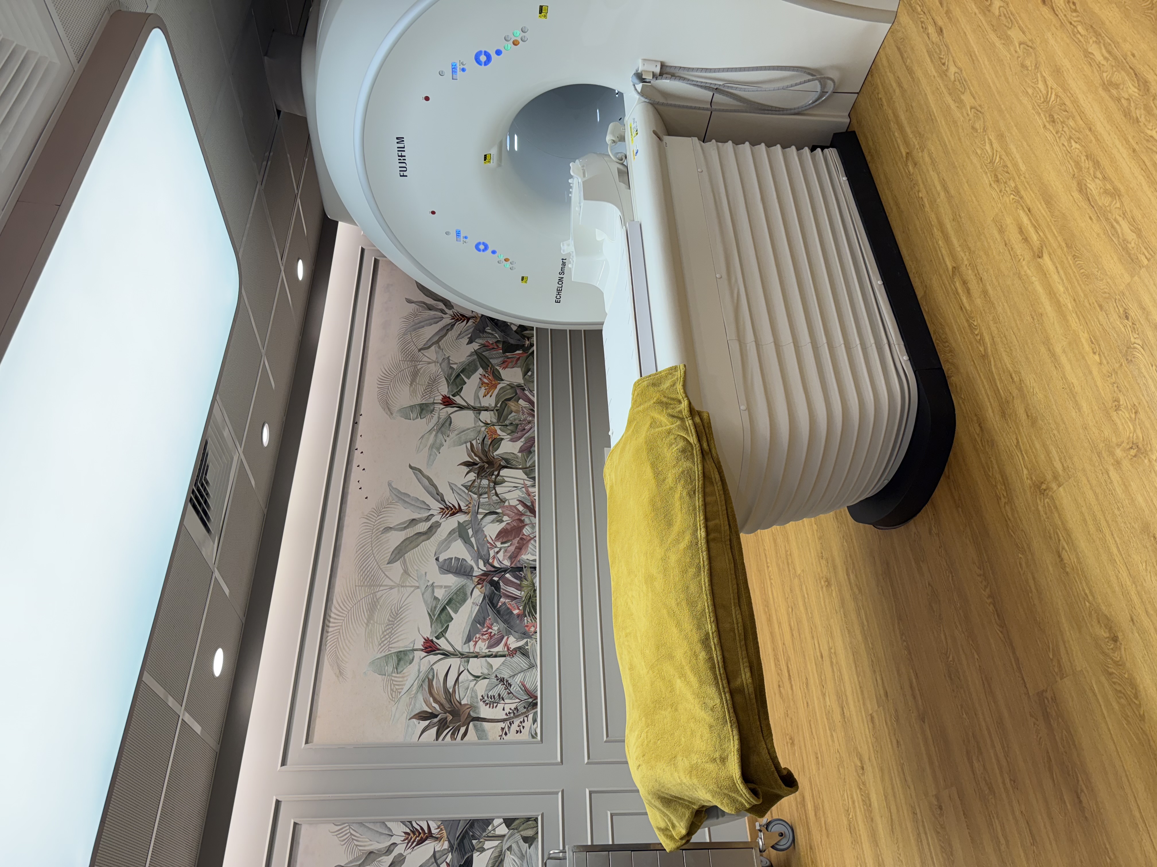
| Scanner | Fujifilm ECHELON Smart 1.5T MRI, with in-scanner music |
|---|
| Reported by | Our radiologists, Dr. Avinash Bansal and Dr. Vaishnavi Bansal |
|---|
| Report | Most reports the same day, sent to your WhatsApp |
|---|
| Price | Exact price confirmed on WhatsApp before your visit |
|---|
| Hours | Mon-Sat 8am-10pm, Sun 8am-5pm; ground floor, step-free entry |
|---|
What an ankle and foot MRI shows
MRI uses a strong magnet and radio waves, not X-rays. It shows ligaments, tendons, cartilage and swelling inside the bone, so it can find an injury when the X-ray looks normal. In Hindi this is टखने का MRI, and in Marathi घोट्याचा MRI. It is used to look for:
- Ligament sprains and tears: most often the ATFL (anterior talofibular ligament) on the outer side, and also the deltoid ligament on the inner side and the syndesmosis joining the two leg bones just above the ankle (a "high ankle sprain")
- Achilles tendon thickening (tendinopathy) and partial or complete tears
- Peroneal tendon injuries on the outer ankle, and tibialis posterior tendon injuries on the inner side, which can cause a flat foot in adults
- Osteochondral lesions: damage to the cartilage and underlying bone of the talus, a common cause of deep ankle pain that continues after a sprain
- Bone bruises, hidden fractures, and stress fractures of the metatarsals, heel bone or navicular
- Plantar fasciitis (a thickened, irritated plantar fascia) and other causes of heel pain
- Morton's neuroma: a thickened nerve at the base of the toes, usually between the third and fourth, causing burning pain in the ball of the foot
- Impingement (pinching) at the front or back of the ankle, arthritis, ganglion cysts and other lumps
- In diabetes: bone infection (osteomyelitis), a pocket of pus (abscess), and Charcot foot, where the bones and joints of a foot with reduced feeling weaken and can collapse
When doctors ask for an ankle or foot MRI
Orthopaedic surgeons, sports medicine doctors, rheumatologists and diabetologists commonly request it for:
- A sprain that is still painful, swollen or "giving way" several weeks later, despite rest and physiotherapy
- A suspected hidden fracture or stress fracture when the X-ray is normal
- Heel or Achilles pain that has not settled with treatment, or a suspected Achilles tear
- Burning pain or numbness between the toes, or a lump in the foot
- Planning ankle or tendon surgery, or checking the ankle afterwards
- A non-healing diabetic foot ulcer or a hot, swollen foot, to see whether the bone is involved
Go to a hospital emergency department first if, after an injury, you cannot put any weight on the foot, the ankle looks deformed, a bone has broken the skin, the foot turns cold, pale or numb, or severe pain keeps getting worse despite painkillers. A sudden "snap" at the back of the ankle may be a torn Achilles tendon: see an orthopaedic doctor the same day. If you have diabetes and your foot becomes hot, red or swollen, keep weight off it and see your doctor urgently; with fever, pus or spreading redness, go to an emergency department. A painful, swollen calf may be a blood clot: see a doctor the same day for a
venous Doppler, not an MRI, and go to an emergency department at once if you also have chest pain or breathlessness.
X-ray, ultrasound or MRI: which comes first?
An MRI is not always the first test. Your doctor chooses based on the question:
- X-ray usually comes first after a twist or fall to check for a fracture, and for arthritis, bunions and a diabetic foot. Tell us before any X-ray if you are, or might be, pregnant.
- MSK ultrasound (sonography, USG) is often enough for plantar fasciitis, the Achilles tendon, Morton's neuroma and lumps near the surface, with no radiation.
- MRI is used when these tests have not answered the question, or for deeper problems: ligaments and cartilage inside the ankle, bone bruises, stress fractures, bone infection, or planning surgery.
How to prepare
Most ankle and foot MRIs need no special preparation: eat, drink and take your regular medicines as normal. If contrast is planned, our team will tell you whether to avoid food for a few hours beforehand. Our team confirms the exact preparation when you book on WhatsApp.
- Tell us before booking about a pacemaker, cochlear implant, nerve stimulator, brain surgery clip or any other implant. Some pacemakers and MR-conditional implants can be scanned under supervision, following the device's own conditions; others cannot.
- Insulin pumps, glucose sensors and medicine patches worn on the skin usually must come off before you enter the scanner room: tell us, and bring a spare or plan with your doctor.
- Tell us if you have ever had metal in your eye, worked with metal (grinding or welding), or have shrapnel or metal fragments in the body.
- Wear loose clothes without metal (a gown is available), and remove anklets, toe rings and other jewellery.
- If your ankle is in a plaster cast, walking boot or external metal frame (fixator), tell us when you book so our team can advise you.
- Bring your prescription and any earlier X-ray, ultrasound or MRI reports of the same foot.
What happens during the scan
You lie on your back with the ankle or foot in a padded coil and go into the scanner feet first, so your head usually stays near the opening or outside it. The scanner makes knocking sounds; the Fujifilm ECHELON Smart is designed to reduce this noise, and in-scanner music is available. Please keep the foot still, as movement blurs the pictures.
The scan usually takes about 20-40 minutes for one ankle or foot, longer if contrast is given. If you are very anxious in closed spaces, or the scan is for a young child, tell us when you book: sedation is available when needed, under the supervision of an anaesthesiologist. Sedation needs a period without food, which our team will explain, and afterwards the patient needs an adult to take them home and should not drive that day. Without sedation, you can leave as soon as the scan is done.
Safety, contrast and pregnancy
MRI uses no radiation, and most ankle and foot MRIs are done without contrast. An injection of contrast (gadolinium) into an arm vein may be needed when infection, a lump or joint inflammation is being assessed, including many diabetic foot scans. A recent kidney function (creatinine) report may then be requested; if you do not have one, the test can be done at the centre before the scan. Tell us about allergies, any earlier reaction to contrast, asthma, kidney disease, and diabetes medicines such as metformin. If you are breastfeeding, mention it; stopping is usually not necessary after this type of contrast.
If you are, or might be, pregnant, tell us before booking. Contrast is generally avoided in pregnancy, and your doctor and the radiologist will decide whether the scan should go ahead or wait.
Ankle and foot MRI at AV Scan, Dadar West
Ankle and foot MRIs are reported by our radiologists, Dr. Avinash Bansal and Dr. Vaishnavi Bansal; Dr. Avinash Bansal, MD (Radiology), has particular expertise in musculoskeletal (MSK) imaging. The report describes the ligaments, tendons, cartilage and bones, including whether any tear is partial or complete and where it is, so your orthopaedic doctor or physiotherapist can plan treatment. Most reports the same day, sent to your WhatsApp.
The exact price is confirmed on WhatsApp before your visit. Ground Floor (step-free entrance), Shilpa Apartment, Shankar Ghanekar Marg, Dadar West / Prabhadevi, Mumbai 400028, near Shivaji Park and Siddhivinayak Temple. WhatsApp or call +91 98200 51511, or book online.
- Step-free ground-floor entry, helpful if you are on crutches or in a walking boot
- Cashless with most major insurers and TPAs; empanelled for CGHS, ECHS and MJPJAY
- Open Monday to Saturday 8am-10pm and Sunday 8am-5pm
- Other joint scans here include knee MRI and shoulder MRI; see all MRI scans