At a Glance
| Also called | MSK USG, USG shoulder, tendon or joint sonography |
|---|
| Radiation and contrast | None; sound waves, no injection |
|---|
| Preparation | Usually none; wear loose clothing (confirmed on WhatsApp) |
|---|
| Scan time | Usually about 15-30 minutes for one area |
|---|
| Performed and reported by | Dr. Avinash Bansal, MD (Radiology) |
|---|
| Report | Most reports the same day, sent to your WhatsApp |
|---|
| Pregnancy | Safe; no radiation |
|---|
| Price | Exact price confirmed on WhatsApp before your visit |
|---|
| Hours | Mon-Sat 8am-10pm, Sun 8am-5pm |
|---|
What an MSK ultrasound shows
High-frequency sound waves make live pictures of the parts that move your joints: tendons, muscles, the fluid-filled cushions called bursae, many ligaments and the bone surface. Patients also call it MSK USG or shoulder sonography (कंधे की सोनोग्राफी / खांद्याची सोनोग्राफी). It shows these well:
- Shoulder: rotator cuff tendinopathy and tears (partial or full-thickness), bursitis, calcium deposits (calcific tendinitis) and the biceps tendon
- Elbow: tennis elbow (outer side), golfer's elbow (inner side) and a swollen bursa at the point of the elbow
- Wrist and hand: De Quervain's tenosynovitis (thumb-side wrist pain), trigger finger and ganglion cysts
- Knee: the patellar and quadriceps tendons, and a Baker's cyst behind the knee
- Heel and ankle: plantar fasciitis, Achilles tendon thickening (tendinopathy) or tears, and the ankle tendons
- Muscles: tears after sport or a fall, and collections of blood (haematoma)
- Lumps: whether a lump is fluid-filled or solid, its size, and its blood flow on colour Doppler
- Joints: fluid in the joint (effusion) and swelling of the joint lining (synovitis). Ultrasound cannot tell whether the fluid is infected; a sample has to be tested for that.
Why ultrasound: live movement and comparison with the other side
Ultrasound can be done while you move, which a standard X-ray or MRI cannot. Dr. Avinash Bansal can watch the rotator cuff glide under the bony roof of the shoulder as you lift your arm, see a trigger finger catch as you bend it, press gently exactly where it hurts, and check your other side to see what is normal for you. With no radiation, it can be repeated to check healing, and it helps if you cannot have an MRI (some pacemakers or implants, or severe claustrophobia).
What ultrasound cannot show: when an MRI or X-ray is needed
Sound waves do not pass through bone, so ultrasound cannot see deep inside a joint. Joint cartilage, the bone marrow (bone bruising or an early stress fracture), the labrum of the shoulder and the ligaments and menisci inside the knee, such as the ACL, need an MRI. Dr. Avinash Bansal also reports shoulder and knee MRI, done on the Fujifilm ECHELON Smart 1.5T MRI at the centre.
For a suspected fracture or arthritis, an X-ray is usually the first step. Ultrasound shows whether a lump is solid, but cannot always show that a solid lump is harmless: a lump that is deep, larger than a golf ball, growing or painful usually needs an MRI as well. If the scan cannot fully answer your doctor's question, the report says so and suggests the next test.
When doctors request an MSK ultrasound
Orthopaedic surgeons, rheumatologists, sports medicine doctors and family doctors commonly request it for:
- Shoulder pain when lifting the arm overhead, reaching behind your back or lying on that side at night
- Weakness lifting the arm after a fall or a heavy lift (after a fall, an X-ray is often done first)
- Elbow pain when gripping, pain on the thumb side of the wrist, or a finger that clicks or locks
- Heel pain that is worst with the first steps in the morning
- Pain or swelling at the back of the ankle, or a sudden calf or thigh pain during sport
- A lump near a joint or tendon, or a swollen joint
Go to a hospital emergency department first if, after an injury, you cannot bear weight or move the joint, the limb looks deformed, or pain keeps getting worse in a tight, swollen limb, or if a joint is suddenly hot, red and swollen with fever. A sudden snap at the back of the ankle may be a torn Achilles tendon: see an orthopaedic doctor the same day. A painful, swollen calf, especially after surgery, a long journey or bed rest, may be a blood clot (DVT) rather than a muscle tear: see a doctor urgently, as the test for a clot is a venous Doppler scan, not an MSK ultrasound. Go to an emergency department at once if you also have chest pain or breathlessness.
How to prepare
Very little preparation is needed. Our team confirms the exact preparation when you book on WhatsApp.
- No fasting or full bladder; take your usual medicines.
- Wear loose clothing that makes the painful area easy to reach, such as a sleeveless T-shirt for the shoulder. A gown is available if needed.
- Remove rings, bangles and watches for a hand or wrist scan.
- Bring your prescription and any previous X-ray, MRI or ultrasound reports and CDs.
- Tell us about any surgery or steroid injection in the area, and roughly when, as both can change how the tissues look.
What happens during the scan
For a shoulder, elbow or wrist you usually sit on a stool; for a knee, heel or ankle you lie on the couch. Dr. Avinash Bansal performs the scan himself: he asks where it hurts and which movements bring the pain on, then moves a small handheld probe over the skin with gel. He may ask you to lift your arm or bend a finger, press over the sore spot, or scan the other side; if anything is uncomfortable, tell him and he will pause.
No sedation is needed. It usually takes about 15-30 minutes for one area, and you can drive and return to work straight afterwards.
Safety and contrast
MSK ultrasound uses no radiation, no injection and no contrast dye, and it is safe in pregnancy. If your doctor later advises an MRI or CT: most shoulder and knee MRIs do not need contrast, and when contrast is needed, a recent kidney function (creatinine) report may be requested; please tell us about any allergies and any diabetes medicines such as metformin. Before any CT, X-ray or MRI, tell us if you are, or might be, pregnant, and before any MRI, tell us about a pacemaker or any other implant.
Your report, and if you need a procedure
The report describes each structure examined, with the size and position of any tear, inflammation, calcium or fluid, so your doctor or physiotherapist can plan treatment. Most reports the same day, sent to your WhatsApp.
If a lump does not look clearly harmless, your doctor may advise an MRI, a needle sample taken under ultrasound guidance (biopsy or FNAC), or a specialist opinion before any needle test, depending on the lump. If your doctor has advised an ultrasound-guided procedure, ask our team on WhatsApp whether it can be done at the centre, and tell us if you take blood thinners.
MSK ultrasound at AV Scan, Dadar West
Ground Floor (step-free entrance), Shilpa Apartment, Shankar Ghanekar Marg, Dadar West / Prabhadevi, Mumbai 400028, near Shivaji Park and Siddhivinayak Temple. WhatsApp or call +91 98200 51511, landline 022-3522 0711, or book online.
- Performed and reported by Dr. Avinash Bansal, MD (Radiology), whose areas of work include MSK imaging
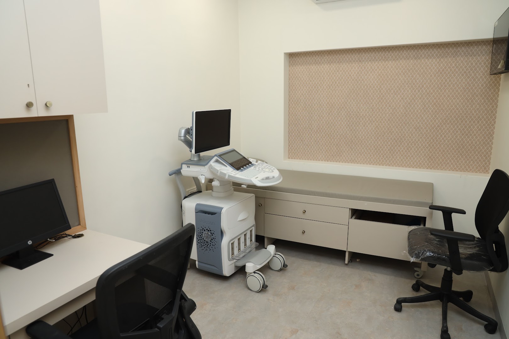
- Ultrasound systems at the centre: GE Voluson E10 and Philips Affiniti 70G
- Digital X-ray and a 1.5T MRI at the same centre if your doctor advises a further test
- The exact price is confirmed on WhatsApp before your visit
- Cashless with most major insurers and TPAs; empanelled for CGHS, ECHS and MJPJAY
- Open Monday to Saturday 8am-10pm and Sunday 8am-5pm