At a Glance
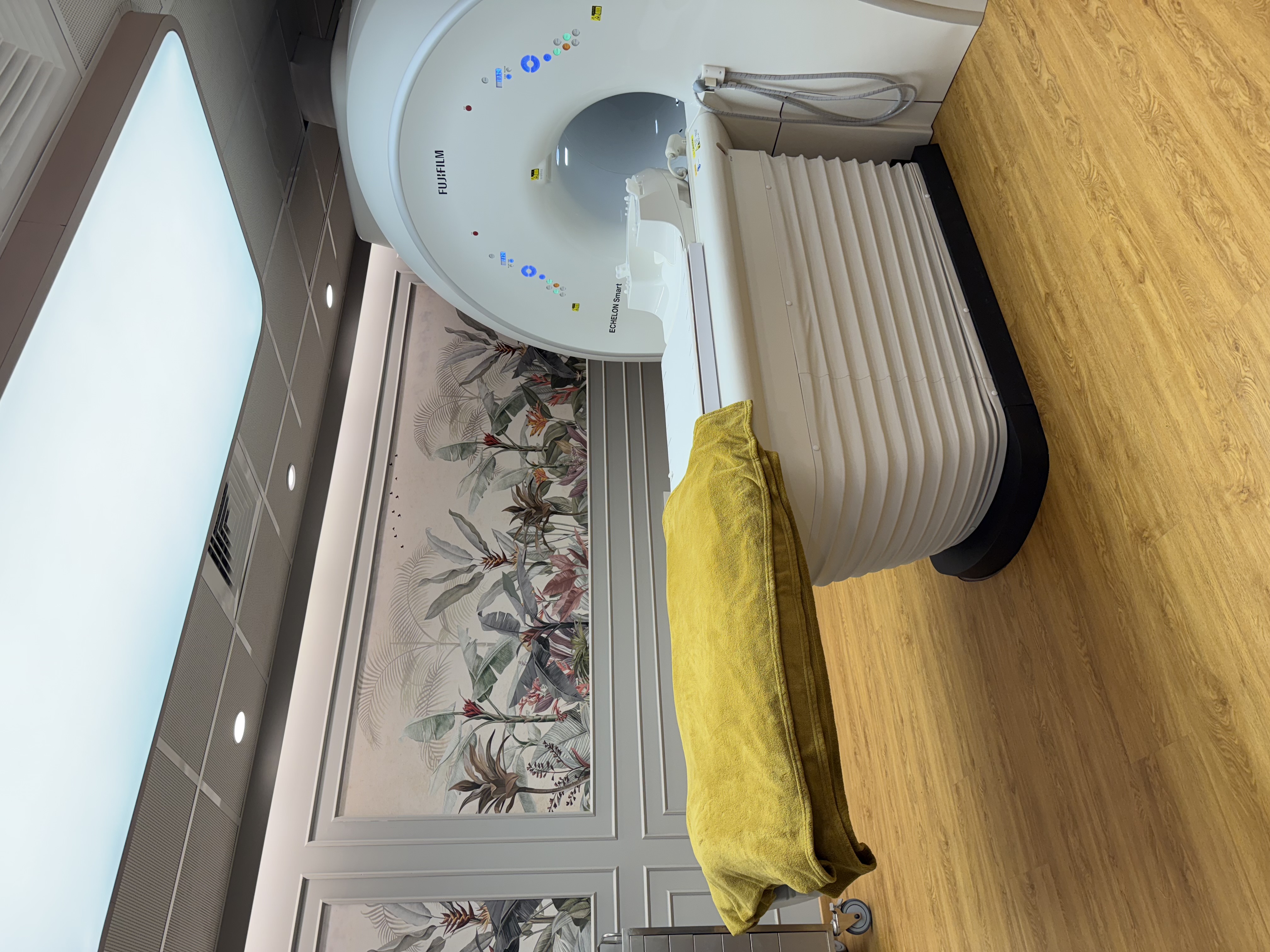
| MRI | Fujifilm ECHELON Smart 1.5T, 70 cm wide bore, quieter scanning, in-scanner music |
|---|
| CT | Fujifilm Supria CT with Intelli IP iterative reconstruction |
|---|
| Mammography | Fujifilm Amulet Innovality 3D mammography (tomosynthesis), paired with breast ultrasound |
|---|
| Ultrasound | GE Voluson E10 and Philips Affiniti 70G, with colour Doppler |
|---|
| Bone density | DEXA for bone density and whole-body composition |
|---|
| X-ray | Digital X-ray; walk-ins welcome with a doctor's prescription |
|---|
| Heart and lungs | 2D echo, ECG, TMT and PFT |
|---|
| Post-processing | Fujifilm Synapse 3D for complex CT and MRI studies |
|---|
| Imaging reported by | Dr. Avinash Bansal and Dr. Vaishnavi Bansal, MD (Radiology), FMF (UK) certified |
|---|
| Open | Mon-Sat 8am-10pm, Sun 8am-5pm; ground floor, step-free entry |
|---|
MRI: Fujifilm ECHELON Smart 1.5T with a 70 cm wide bore
MRI uses a strong magnet and radio waves, not X-rays, to show soft tissue in detail: the brain, spinal cord and discs, ligaments, cartilage and the organs of the abdomen and pelvis. We use it for brain, spine and joint MRI, MRCP, and breast, prostate, cardiac and fetal MRI; see all MRI scans.
The magnet is always on, so safety comes first. Tell us before booking about a pacemaker or defibrillator, a cochlear implant, a brain aneurysm clip, an insulin pump or glucose sensor, any other implant, or metal fragments in the eye (for example from welding or grinding work). You fill in a safety checklist before the scan and remove all metal, including jewellery, hairpins and medicine patches. Tell us if you are or could be pregnant. Contrast (a gadolinium injection) is needed for some questions, such as a suspected tumour, infection or inflammation; if it is planned, a recent creatinine (kidney function) report may be requested; mention allergies, any earlier reaction to contrast, kidney disease and diabetes medicines. Our team confirms the exact preparation when you book on WhatsApp; see the MRI preparation guide.
- 70 cm wide bore: room around your shoulders and face, which helps if you are broad-built or feel closed in. It is still a tunnel, open at both ends.
- Quieter scanning: the scanner is designed to reduce noise, and you are given ear protection and in-scanner music. It still knocks while it takes pictures.
- Sedation is available when needed, under the supervision of an anaesthesiologist. You will need to fast beforehand and have an adult take you home; our team gives you the instructions.
CT: Fujifilm Supria CT with Intelli IP
CT uses X-rays and a computer to make thin cross-sectional pictures of the body. It is commonly used for the lungs (HRCT chest), kidney stones (CT KUB), sinuses (HRCT PNS), the head, and contrast (CECT) scans of the abdomen, including triple-phase CT. CT urography, CT angiography, calcium scoring and CT-guided biopsy are also done here; see all CT scans.
- Intelli IP, Fujifilm's iterative reconstruction software, reduces image noise, which helps keep the radiation dose lower while the pictures stay clear. Each scan is planned to use as little radiation as is needed to answer the clinical question (the ALARA principle, as low as reasonably achievable); see CT radiation explained.
- A short ring, not a long tunnel; the scan itself usually takes from a few seconds to a few minutes.
- Contrast (CECT) is used when blood vessels or organs such as the liver, pancreas or kidneys need to be shown more clearly. For a contrast scan: about 4 hours without food, water allowed. A recent creatinine report may be requested (if you have none, the test can be done at the centre before the scan). Tell us about allergies, asthma, kidney disease or any earlier reaction to contrast; if you take metformin, we check your kidney function (eGFR) and advise you.
- Tell us first if you are or could be pregnant. Our team confirms the exact preparation on WhatsApp.
Sudden weakness of the face, arm or leg, slurred speech, a sudden severe headache or a serious head injury: go to a hospital emergency department first, not a scan centre.
3D mammography: Fujifilm Amulet Innovality, paired with breast ultrasound
The Fujifilm Amulet Innovality performs digital breast tomosynthesis (3D mammography). While the breast is compressed for several seconds for each view, the X-ray tube moves in a short arc and takes a series of low-dose images that are built into thin slices, so the radiologist can look through the breast layer by layer. Overlapping tissue can then be less likely to hide a problem or to look like one, which helps particularly when breast tissue is dense. No single test finds every cancer, which is one reason ultrasound is added.
Every mammogram is paired with a breast ultrasound in the same visit, performed by Dr. Vaishnavi Bansal herself, our breast imaging specialist, and same-day ultrasound-guided biopsy is possible when needed. Avoid deodorant, talcum powder and perfume on the day, and tell us if you are pregnant or breastfeeding or have breast implants; our team confirms the preparation on WhatsApp. See 3D mammography, breast ultrasound and biopsy and FNAC.
A new breast lump, nipple discharge or skin change should be checked promptly even if a recent mammogram was normal. WhatsApp us or see your doctor.
Ultrasound (sonography): GE Voluson E10 and Philips Affiniti 70G
Ultrasound uses sound waves, not X-rays, and has no known harmful effects when used for a medical reason, which is why it is the main scan used in pregnancy. The GE Voluson E10 is designed for pregnancy and women's health imaging, including 3D. The Philips Affiniti 70G is a general-purpose system for the abdomen, thyroid and neck, blood vessels, muscles and tendons. Both have colour Doppler, which shows blood flow.
Pregnancy scans and fetal echo are done by our radiologists, Dr. Avinash Bansal and Dr. Vaishnavi Bansal, both FMF (UK) certified. See also ultrasound, colour Doppler and MSK ultrasound. In Hindi and Marathi it is called सोनोग्राफी (sonography); prescriptions often write USG. Some scans need a full bladder or fasting, which our team confirms when you book on WhatsApp.
DEXA: bone density and body composition
A DEXA scan uses very low-dose X-rays at two different energies to measure bone density at the spine, hips and, when needed, the forearm; it is the test doctors use to diagnose osteoporosis and to follow its treatment. It also measures whole-body composition: fat mass, lean (muscle) mass and where they sit. You lie on an open table while the scanner arm passes over you. Tell us if you are or could be pregnant, or if you have recently had a barium test, a contrast scan or a nuclear medicine scan, as these can affect the reading. You may be asked not to take calcium tablets for 24 hours before the scan and to wear clothes without metal; our team confirms any preparation when you book on WhatsApp.
Digital X-ray
Digital X-ray covers the chest, spine, joints, sinuses and abdomen. The images appear on screen within moments, so the radiographer can check them and repeat a view before you leave if needed. The dose is low; tell us first if you are or could be pregnant. Walk-ins are welcome with a doctor's prescription, and plain X-rays usually need no preparation. An X-ray IVU is available when a doctor asks for it; because it uses a contrast injection, it needs booking, and the same kidney function, allergy and metformin checks as a CT with contrast apply. Our team confirms the exact preparation when you book on WhatsApp.
Heart and lung tests: 2D echo, ECG, TMT and PFT
A 2D echo is an ultrasound of the heart's pumping, chambers and valves. An ECG records the heart's electrical activity and usually takes a few minutes. A TMT (treadmill test) records your ECG and blood pressure while you walk at increasing speed and slope. A PFT (spirometry) measures how much air you breathe out and how fast, for asthma, COPD and other breathing problems. For a TMT, wear walking shoes and avoid a heavy meal for a couple of hours before. Do not stop any heart medicine or inhaler on your own; our team tells you whether anything should be paused, on your doctor's advice, and confirms the preparation for each test when you book on WhatsApp. See 2D echo and ECG, TMT and PFT.
Chest pain or pressure happening now, sudden breathlessness or fainting needs a hospital emergency department first, not a TMT or an echo.
Synapse 3D: post-processing for complex scans
Fujifilm Synapse 3D is software our radiologists use on the images the CT and MRI scanners have already taken. For complex studies, such as CT angiography or scans to plan surgery, it builds 3D views of blood vessels, bones and organs and helps with measurements. It adds no time in the scanner and no extra radiation.
Who reads your scans, and visiting us
The equipment takes the pictures; a named radiologist plans the scan and reads it.
- Imaging scans are reported by our radiologists, Dr. Avinash Bansal (musculoskeletal imaging, fetal medicine and Doppler) and Dr. Vaishnavi Bansal (breast imaging), both MD (Radiology) and FMF (UK) certified.
- Most reports the same day, sent to your WhatsApp.
- WhatsApp your prescription; we confirm the scan, preparation and exact price before your visit.
- Cashless with most major insurers and TPAs; empanelled for CGHS, ECHS and MJPJAY (see insurance and TPA).
- Open Monday to Saturday 8am-10pm and Sunday 8am-5pm, on the ground floor with step-free entry, near Shivaji Park and Siddhivinayak Temple (see how to find us).