At a Glance
| Also called | MRI breast, bilateral breast MRI, contrast-enhanced (DCE) breast MRI, breast implant MRI |
|---|
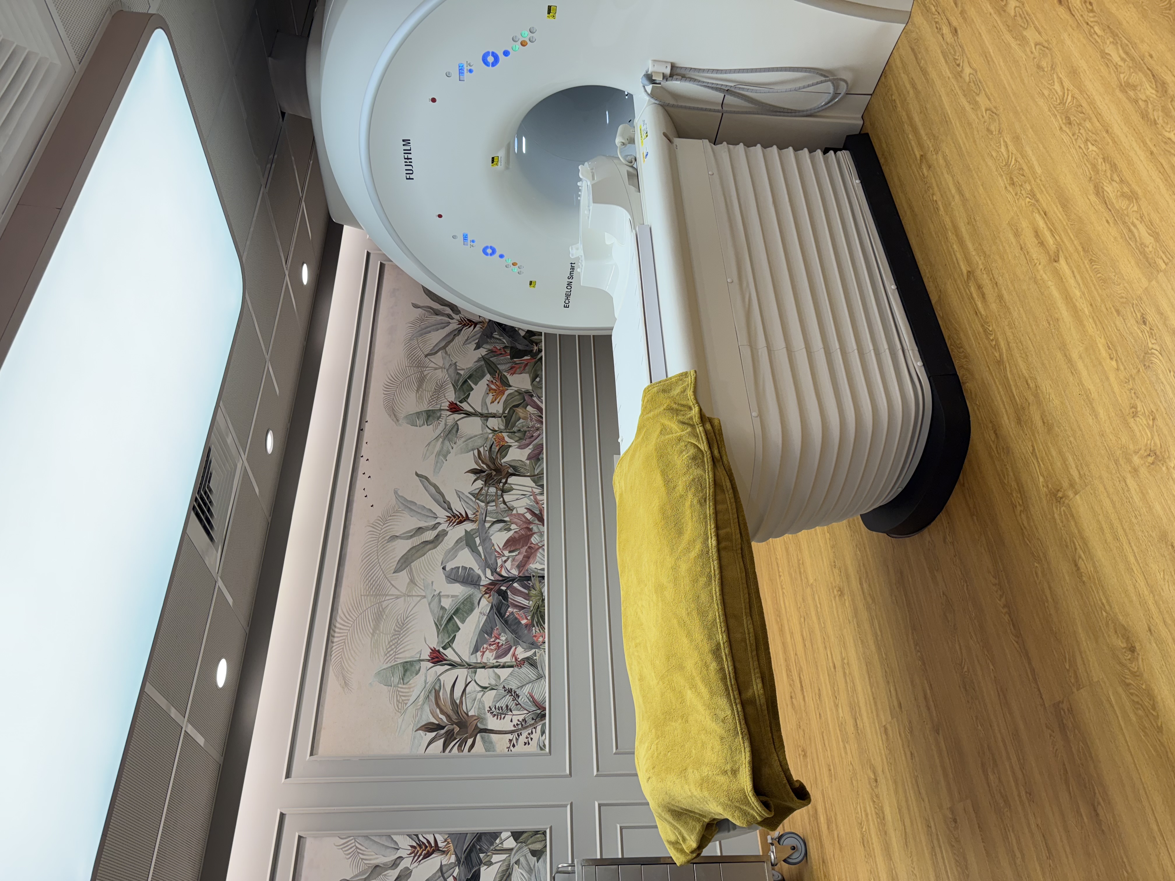
| Scanner | Fujifilm ECHELON Smart 1.5T MRI, with quieter scanning and in-scanner music |
|---|
| Reported by | Dr. Vaishnavi Bansal, MD (Radiology), FMF (UK) certified, breast imaging specialist |
|---|
| Radiation | None (MRI uses a magnet and radio waves) |
|---|
| Contrast | A gadolinium injection for most breast MRIs; usually not needed for an implant-only check |
|---|
| Position | Lying face-down on a padded support |
|---|
| Timing | If you still have periods, often about days 7-14 of your cycle; our team confirms |
|---|
| Time in the scanner | Usually about 20-40 minutes, plus time to change and get positioned |
|---|
| Preparation | Our team confirms the exact preparation when you book on WhatsApp |
|---|
| Report | BI-RADS category; most reports the same day, sent to your WhatsApp |
|---|
| Price | Exact price confirmed on WhatsApp before your visit |
|---|
| Payment | Cashless with most major insurers and TPAs; empanelled for CGHS, ECHS and MJPJAY |
|---|
| Hours and access | Mon-Sat 8am-10pm, Sun 8am-5pm; ground floor, step-free |
|---|
What a breast MRI shows
Breast MRI uses a magnet and radio waves, not X-rays. A contrast dye (gadolinium) is usually injected into an arm vein during the scan; many cancers take it up quickly, so they stand out even in dense breast tissue. Patients also call it MRI breast (स्तन का MRI स्कैन / स्तनाचा MRI स्कॅन). It can show:
- A cancer hidden in dense tissue that a mammogram or ultrasound could not clearly show
- The extent of a known cancer: its size, any other spots in either breast, and whether it reaches the chest wall
- Silicone implant rupture, including a "silent" rupture that cannot be felt
- How a cancer is responding to chemotherapy given before surgery
- Scar tissue versus possible recurrence after breast surgery or radiotherapy
When doctors request a breast MRI
Breast MRI is usually a second step after a 3D mammogram and a breast ultrasound (sonomammography), requested by your surgeon, oncologist or gynaecologist. Common reasons include:
- A finding that is still uncertain after mammography and ultrasound
- A newly diagnosed breast cancer, before the surgeon plans the operation
- Before, during and after chemotherapy given before surgery
- A cancer found in an armpit lymph node when the mammogram and ultrasound look normal
- Yearly screening for women at high risk, as advised by their doctor (for example a BRCA1 or BRCA2 gene change, a strong family history, or radiotherapy to the chest before the age of 30); screening often starts at a younger age than routine mammography, and is usually combined with mammography
Breast MRI for silicone implants
MRI shows the implant shell and the silicone around it in detail, and can find a rupture that causes no change you can see or feel. An implant-only check usually needs no contrast and no particular day of the cycle. A saline implant that ruptures usually goes flat within days, so MRI is used mainly for silicone implants. If the question is cancer, contrast is used, and MRI can assess breast tissue that implants partly hide on a mammogram. New swelling or fluid around an implant years after surgery is usually checked first with ultrasound, and fluid may need to be sampled for testing.
How to prepare for a breast MRI
Our team confirms the exact preparation when you book on WhatsApp. As a general guide:
- Cycle timing: if you still have periods, a contrast breast MRI is often booked for about days 7-14 of your cycle (day 1 is the first day of bleeding), when normal breast tissue takes up less dye. If your cycles are irregular, tell us. A scan to plan cancer treatment is usually not delayed for this.
- Tell us if you take hormone replacement therapy (HRT) or other hormone tablets; do not stop any medicine on your own.
- Food: our team tells you whether to avoid food for a few hours before a contrast scan; take regular medicines as usual unless told otherwise.
- Bring all earlier mammograms, ultrasounds and MRIs (films, CDs or links), biopsy and operation reports, chemotherapy dates and any implant card, for comparison.
- Wear a two-piece outfit; you will change into a gown and remove jewellery, hairpins, underwired bras, watches and hearing aids. Tell us about any medicine skin patch, as some contain metal and must be removed.
- Tell us if lying face-down with your arms raised is difficult for you, or if closed spaces make you anxious.
What happens during the scan
After a metal-safety checklist, a small cannula is placed in an arm vein if contrast is planned. You lie face-down on a padded support with openings for the breasts, which holds the receiver coil. You are given ear protection and a call bell to hold throughout, and the Fujifilm ECHELON Smart has quieter scanning and in-scanner music.
When contrast is used, it is given through the cannula partway through the scan while you stay still, and the pictures taken before and after it are compared. The scan usually takes about 20-40 minutes, plus time to change and get positioned. Afterwards you can usually eat, drive and go back to work straight away. If you had sedation, an adult must take you home, and you should not drive or make important decisions for the rest of the day.
Contrast and MRI safety
MRI contrast (gadolinium) is different from the iodine dye used for CT. Reactions are uncommon and usually mild, and our team is prepared to manage them. A recent kidney function (serum creatinine) report may be requested; if you do not have one, the test can be done at the centre before your scan. Tell us about any allergy or previous reaction to MRI or CT contrast, asthma, kidney disease or dialysis, diabetes and diabetes medicines such as metformin (we check your kidney function and advise you), and whether you are, or might be, pregnant, or are breastfeeding.
Before booking, tell us about a pacemaker or defibrillator, cochlear implant, nerve stimulator, brain aneurysm clip, metal fragments in the eye, a chemotherapy port, or any other implant. Some pacemakers and MR-conditional implants can be scanned under supervision, following the device's conditions; others cannot be scanned at all. Most breast tissue expanders placed after mastectomy have a magnetic valve and usually cannot go into the scanner; bring the expander details so we can check. Silicone and saline implants, and the small marker clip left after a breast biopsy, are generally not a problem.
If closed spaces make you very anxious, sedation is available when needed, under the supervision of an anaesthesiologist; our team gives you separate instructions, including fasting, if it is planned.
A breast MRI is a planned test. A hot, red, painful breast with fever, or an implant that suddenly becomes painful and swollen, needs your doctor the same day; if you feel very unwell, go to a hospital emergency department first.
Your report, and why some women need a second look
Dr. Vaishnavi Bansal reports your MRI alongside your earlier mammograms and ultrasounds, using the standard BI-RADS categories. A normal breast MRI is reassuring, but no test finds every cancer; see your doctor about any new lump or change, even if a recent scan was normal.
MRI is very sensitive, so it also picks up harmless spots, and a finding can lead to further tests that turn out normal. If a spot needs a closer look, the next step may be:
- A second-look ultrasound aimed at the exact spot seen on MRI
- An ultrasound-guided biopsy if the spot is found; a same-day biopsy at the centre is possible when needed
- A follow-up MRI, usually after about 6 months, for a probably harmless spot (BI-RADS 3)
- An MRI-guided biopsy if the spot shows only on MRI; your surgeon advises on this
Breast MRI at AV Scan, Dadar West
WhatsApp +91 98200 51511 with a photo of your prescription, call the same number, or use our booking page. Our team confirms the day, the preparation and the exact price before your visit.
- Reported by Dr. Vaishnavi Bansal, MD (Radiology), FMF (UK) certified, who also performs every breast ultrasound here herself
- Fujifilm ECHELON Smart 1.5T MRI, 3D mammography (Fujifilm Amulet Innovality), breast ultrasound and ultrasound-guided breast biopsy at the same centre
- Cashless with most major insurers and TPAs; empanelled for CGHS, ECHS and MJPJAY
- Open Monday to Saturday 8am-10pm and Sunday 8am-5pm; most reports the same day, sent to your WhatsApp
- Ground floor with a step-free entrance, Shankar Ghanekar Marg, near Shivaji Park and Siddhivinayak Temple