At a Glance
| Arthritis or bone injury | X-ray first, usually knee AP and lateral; a skyline view for the kneecap |
|---|
| Tendons, fluid, Baker's cyst | MSK ultrasound (USG knee); no radiation |
|---|
| Ligaments (ACL), meniscus, cartilage, bone bruise | MRI, usually after an orthopaedic doctor has examined the knee; no radiation |
|---|
| Go to emergency first | Cannot stand on the leg after an injury, an obvious deformity, a cold or numb foot, or a hot, swollen knee with fever |
|---|
| Who decides | Your doctor, after examining you |
|---|
| Reported by | Our radiologists, Dr. Avinash Bansal and Dr. Vaishnavi Bansal; musculoskeletal imaging led by Dr. Avinash Bansal |
|---|
| Report | Most reports the same day, sent to your WhatsApp |
|---|
| Price | Exact price confirmed on WhatsApp before your visit |
|---|
| Open | Mon-Sat 8am-10pm, Sun 8am-5pm; ground floor, step-free |
|---|
Warning signs: go to a hospital emergency department first
Do not wait for a scan appointment if you have:
- A knee injury after which you cannot stand or walk on the leg
- A knee or leg that looks deformed, a kneecap that has slipped out of place and stayed out, or a wound over the joint
- A foot that turns cold, pale or numb after a knee injury
- A hot, red, swollen knee with fever or feeling unwell, even without an injury
A hot, swollen knee with fever can be an infected joint, which can damage the cartilage within days. A cold, pale or numb foot after a bad knee injury can mean the main artery behind the knee is injured and needs treatment within hours.
X-ray first: arthritis and bone injury
An X-ray is usually the first test for knee pain that has built up over months or years, especially after about 40, and after a fall or a direct blow. It shows the joint-space narrowing and bony spurs of osteoarthritis, fractures and loose pieces of bone, but not ligaments, the meniscus or cartilage. The usual request is 'X-ray knee AP and lateral' (front and side views) of one or both knees; a skyline view shows the kneecap. In Hindi, knee pain is often called घुटने का दर्द; in Marathi, गुडघेदुखी.
For arthritis, an X-ray is usually all the imaging needed, and with typical symptoms after middle age your doctor may not need any scan to make the diagnosis. A hairline fracture may not show at first; if pain continues, your doctor may repeat the X-ray or ask for an MRI. The dose from a knee X-ray is very small; see digital X-ray.
Ultrasound: tendons, fluid and a Baker's cyst
Knee sonography uses sound waves, not radiation, and shows the structures near the surface: the quadriceps and patellar tendons (including jumper's knee and tendon tears), the ligaments on either side, fluid in the joint and swelling of its lining (synovitis), a swollen bursa from kneeling (housemaid's knee) and a Baker's cyst, a fluid-filled swelling behind the knee. The radiologist can press where it hurts and compare it with your other side.
It cannot see deep inside the joint: the ACL, most of the menisci, the cartilage and the bone marrow need an MRI. It shows fluid in the joint but not whether it is infected or caused by gout; only testing a sample of the fluid can show that. See MSK ultrasound.
MRI: ligaments, meniscus, cartilage and bone bruise
A knee MRI shows the whole joint without radiation: the ACL and PCL, the side ligaments, both menisci (the C-shaped cushions between the thigh and shin bones), cartilage, tendons and bone marrow. It is usually requested after an orthopaedic or sports medicine doctor has examined the knee, for:
- A twisting injury with a 'pop', rapid swelling and a knee that gives way: a possible ACL tear
- Locking, catching or a knee that will not fully straighten: a possible meniscus tear
- A kneecap that slipped out of place, to check the ligament that holds it and the cartilage
- Pain not settling after several weeks of physiotherapy, or before keyhole surgery (arthroscopy)
- Sudden knee pain in an older person with a normal X-ray: possibly a small stress-type fracture in the bone just beneath the cartilage
After a knee injury: when to scan
Most minor sprains, and many aches around the kneecap in active young people, settle within a few weeks with rest, ice and physiotherapy, and need no scan. See a doctor within a few days if the knee swelled up within hours, you felt a pop, or it keeps giving way: rapid swelling often means bleeding inside the joint, as with an ACL tear or a slipped kneecap. A knee that locks and will not straighten needs an orthopaedic opinion within a day or two.
A very swollen, painful knee can be hard to examine at first, so your doctor may re-examine it once the swelling settles, although the MRI itself can be done while it is swollen. Bone bruises, which help confirm a ligament injury, are clearest in the first weeks and usually fade over the following weeks to months. More on our knee MRI page.
Knee pain that may not start in the knee
Hip problems can cause pain felt only in the knee, especially in children, teenagers and older adults, so a doctor may also ask for a hip X-ray. A child or teenager who limps with knee pain should see a doctor promptly; with a fever, go to an emergency department. Pain down the front of the thigh into the knee can come from a pinched nerve in the back; see which scan for back pain.
A burst Baker's cyst can cause calf pain and swelling that looks like a blood clot (DVT), and the two can occur together. A painful, swollen calf needs a doctor urgently; the test for a clot is a venous Doppler scan. With chest pain or breathlessness, go to an emergency department at once.
Preparing, safety and contrast
Our team confirms the exact preparation when you book on WhatsApp. A knee X-ray, ultrasound or plain MRI needs no fasting; take your usual medicines. Wear shorts or loose trousers that roll up above the knee, and bring earlier knee scans and reports, and details of any knee operation.
Tell us before an X-ray if you are or might be pregnant; the knee is far from the womb, so the dose to the baby is extremely small, but we still need to know. Ultrasound uses no radiation.
When you book an MRI, tell us about a pacemaker or any implant, metal fragments in the eye (for example from welding or grinding), and any screws or plates, including from an ACL repair or a knee replacement; bring any device card. Some pacemakers and MR-conditional implants can be scanned here under supervision, following the device's conditions; others cannot. Metal in or near the knee can blur the MRI pictures around it, so after a knee replacement your doctor may choose a different test. Tell us if you are or might be pregnant. You go in feet first, so for most adults the head stays near or outside the scanner. The MRI has in-scanner music and quieter scanning, and sedation is available when needed, under the supervision of an anaesthesiologist. See our MRI preparation guide.
Most knee MRIs need no contrast; it is used only for some problems, such as a suspected infection, a lump or tumour, or inflamed joint lining, and is usually avoided in pregnancy. If contrast is planned, our team tells you whether to fast beforehand, and a recent kidney function (creatinine) report may be requested; if you have none, the test can be done at the centre before the scan. Tell us about kidney disease, allergies or asthma, a past contrast reaction, and diabetes medicines such as metformin; metformin is usually continued for MRI contrast, and our team will advise you.
Your knee scan at AV Scan, Dadar West
X-rays are done only when a doctor has asked for them. For a knee MRI we recommend seeing an orthopaedic doctor first; if you do not have a prescription yet, WhatsApp us and our team will advise you. Send a photo of the prescription and our team confirms the test, the preparation and the exact price before your visit.
- Knee X-ray, MSK ultrasound and MRI at one centre
- Reported by our radiologists, Dr. Avinash Bansal and Dr. Vaishnavi Bansal; musculoskeletal (bone, joint and muscle) imaging is led by Dr. Avinash Bansal
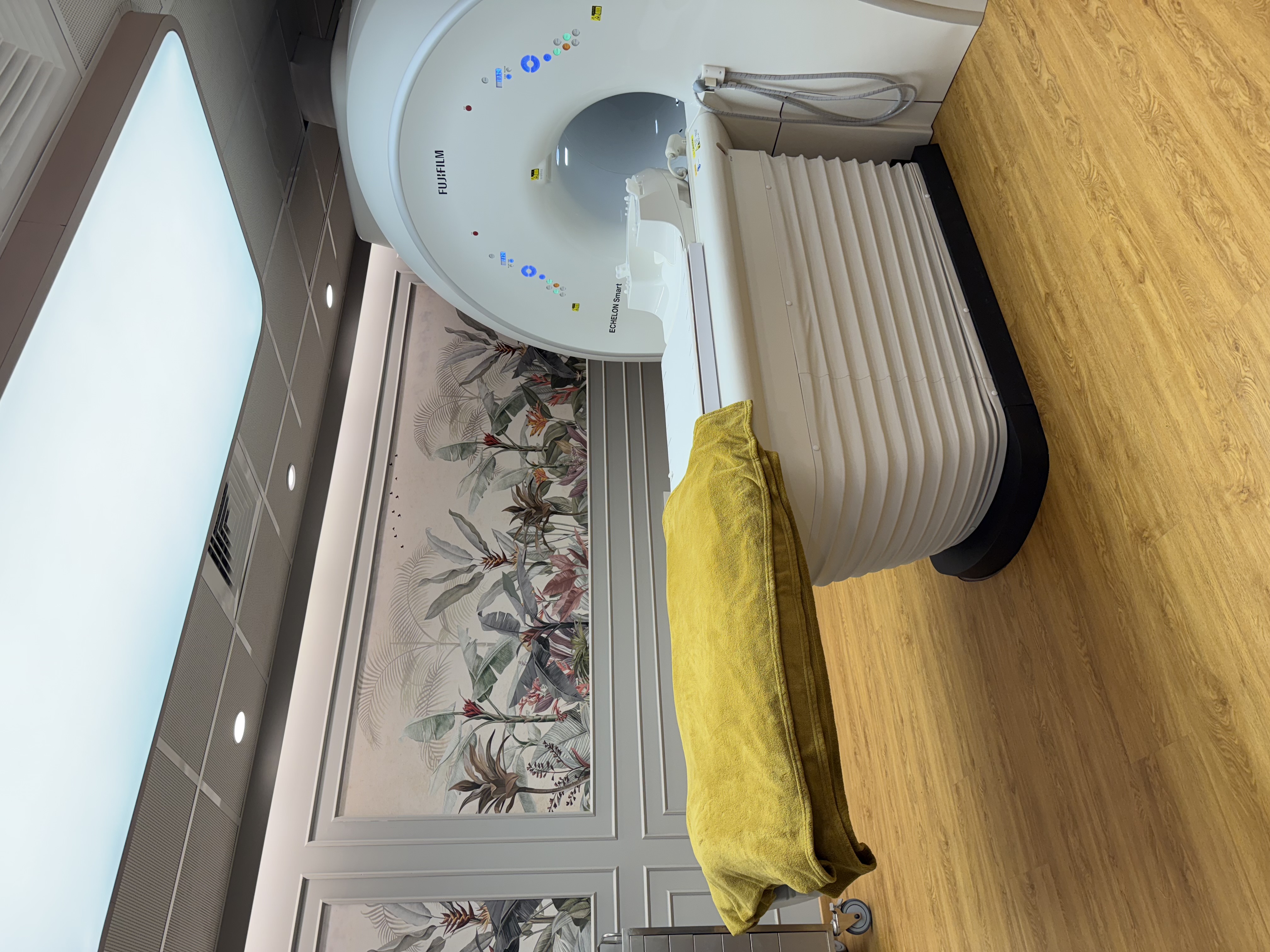
- Fujifilm ECHELON Smart 1.5T MRI; GE Voluson E10 and Philips Affiniti 70G ultrasound; digital X-ray, with walk-ins welcome on a prescription
- Most reports the same day, sent to your WhatsApp
- Cashless with most major insurers and TPAs; empanelled for CGHS, ECHS and MJPJAY (insurance and TPA)
- Open Monday to Saturday 8am-10pm and Sunday 8am-5pm
- Ground floor with step-free entry, near Shivaji Park and Siddhivinayak Temple, which helps when your knee hurts