At a Glance
| Also called | Third-trimester scan, fetal growth scan, obstetric Doppler, growth sonography / USG |
|---|
| Usual timing | Third trimester, often about 28-36 weeks; repeat scans as your obstetrician advises |
|---|
| Performed and reported by | Dr. Vaishnavi Bansal, MD (Radiology), FMF (UK) certified |
|---|
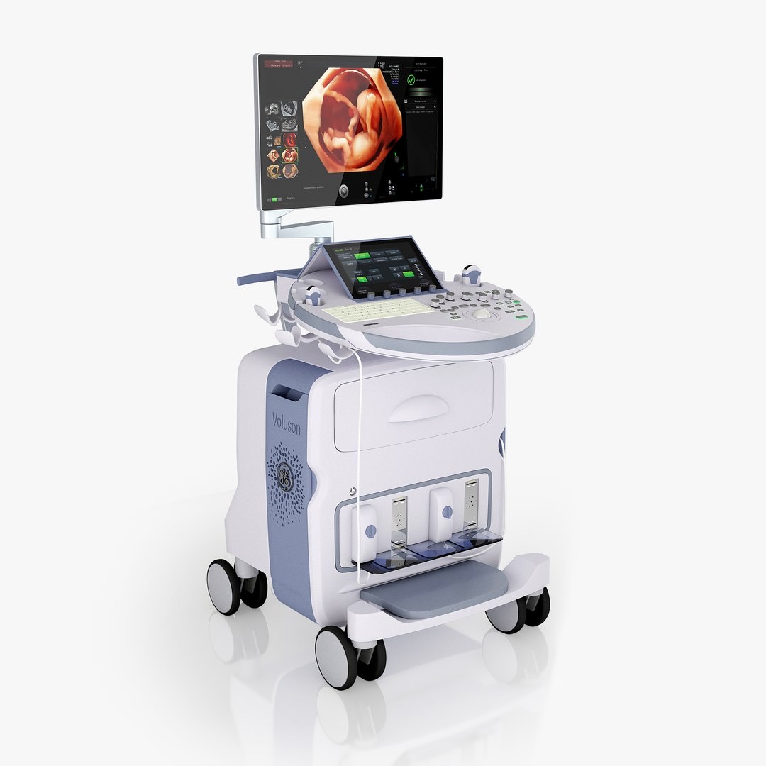
| Ultrasound | GE Voluson E10 |
|---|
| Preparation | No fasting, no full bladder; bring your earlier scan reports |
|---|
| Scan time | Usually about 20-30 minutes |
|---|
| Radiation | None - ultrasound uses sound waves |
|---|
| Report | Most reports the same day, sent to your WhatsApp |
|---|
| Price | Confirmed on WhatsApp before your visit |
|---|
| Hours | Mon-Sat 8am-10pm, Sun 8am-5pm |
|---|
| Access | Ground floor, step-free |
|---|
What a growth scan checks
A growth scan answers a practical question for your obstetrician: is the baby growing as expected, and is the placenta supporting that growth?
- Measurements of the baby's head (BPD and head circumference), tummy (abdominal circumference) and thigh bone (femur length).
- Estimated fetal weight (EFW), compared with the expected range for your weeks of pregnancy. Your weeks come from your early (dating) scan and are not changed by a later scan.
- Growth pattern: today's measurements compared with your earlier scans, because the trend over time says more than a single number.
- Amniotic fluid (liquor), measured as the amniotic fluid index (AFI) or the deepest pocket, to see whether it is low, normal or high.
- Placenta: where it lies, including a recheck if it was low-lying on an earlier scan. If it still looks low, an internal (transvaginal) scan may be advised to measure its distance from the cervix accurately; this is safe in pregnancy and done only with your consent.
- Presentation: whether the baby is head-down (cephalic), bottom-down (breech) or lying across (transverse). Before about 36 weeks many babies still turn on their own.
What the Doppler part adds
Doppler measures blood flow. It helps tell apart a baby who is small but healthy from one held back by the placenta, and helps your obstetrician decide how closely to monitor you. The vessels checked depend on why the scan was requested.
- Umbilical artery: how easily blood flows from the baby into the placenta. Raised resistance suggests the placenta is not working as well as it should. Flow that stops or reverses between heartbeats (absent or reversed end-diastolic flow) is a more serious sign that needs same-day review by your obstetrician.
- Middle cerebral artery (MCA): blood flow in the baby's brain. Low resistance can mean the baby is diverting blood to the brain; with the umbilical artery it gives the cerebroplacental ratio (CPR). A high peak speed can point to anaemia in the baby, for example when the mother has red-cell antibodies (as some Rh-negative mothers do), and this needs specialist fetal medicine care.
- Uterine arteries: the mother's blood supply to the placenta. They may be checked when blood pressure is high or the baby is small, mainly earlier in pregnancy.
- Ductus venosus: a vessel near the baby's heart, checked only for a specific reason, usually when the baby is small early in the third trimester and the umbilical artery Doppler is abnormal. Your obstetrician will then usually arrange close monitoring, often in hospital.
Why your obstetrician may ask for it
- Your bump measures smaller or larger than expected for your weeks.
- A previous baby was born small, or a previous pregnancy had complications.
- High blood pressure or pre-eclampsia, or a raised risk of these on early pregnancy screening.
- Diabetes before or during pregnancy (gestational diabetes), which can make a baby grow large or increase the fluid.
- Reduced fetal movements, after your obstetrician or the labour ward has already assessed you.
- Twin pregnancy, where growth is usually checked more often.
- Too little or too much fluid, or a low-lying placenta, on an earlier scan.
- Other medical conditions in the mother that need closer monitoring.
Do not wait for a scan appointment if your baby is moving less than usual or the pattern of movements has changed. The same applies if you have vaginal bleeding, leaking fluid, a severe headache, blurred vision, sudden swelling of the face or hands, severe tummy pain (including pain under the ribs), or regular tightenings before 37 weeks. Contact your obstetrician or go straight to the labour ward or a hospital emergency department.
How to prepare
Most growth scans need no special preparation. Our team confirms the exact preparation when you book on WhatsApp.
- Eat and drink normally; no fasting or full bladder is needed.
- Bring your obstetrician's prescription, and tell us the week they asked for the scan so it falls at the right time.
- Bring all earlier pregnancy scan reports, especially the dating or NT scan and the anomaly scan.
- Wear a two-piece outfit so your tummy can be uncovered easily.
- Tell us if you are expecting twins, or if your prescription also asks for a fetal heart scan (fetal echocardiography) or another test, so enough time is set aside.
What happens during the scan
You lie on the couch and gel is spread on your tummy. Dr. Vaishnavi moves the probe over your bump to take the measurements, then records the Doppler readings; you may hear a whooshing sound. The scan should not hurt.
It usually takes about 20-30 minutes, longer if the baby's position hides a measurement. Late in pregnancy, lying flat on your back can make you feel faint or sick; tell the team straight away, as turning slightly onto your side usually helps. You are welcome to bring your partner or a family member.
At the end, Dr. Vaishnavi explains the main findings in English, Hindi or Marathi; your obstetrician then decides the plan. If anything needs urgent attention, you will be told before you leave so you can contact your obstetrician without delay.
A growth scan is not a detailed check of the baby's organs, and no scan can detect every problem. Some conditions only become visible later in pregnancy or after birth.
Is it safe?
Yes. Ultrasound uses sound waves, not radiation, and no injection, dye or contrast is used. The Doppler part uses more ultrasound energy than the ordinary picture, so it is used only for a medical reason, at the lowest setting that gives a reliable reading and for as short a time as possible, following international ultrasound safety guidance. Used this way, obstetric Doppler has no known harmful effect on the baby.
Growth scan with Doppler at AV Scan
- Performed and reported by Dr. Vaishnavi Bansal, MD (Radiology), whose FMF (UK) certification includes fetal Doppler. Her work focuses on breast, women's and fetal imaging.
- Scanned on the GE Voluson E10 ultrasound. If your doctor asks for a fetal heart scan, it can be done in the same visit.
- Most reports the same day, sent to your WhatsApp, so you can share them with your obstetrician.
- The exact price is confirmed on WhatsApp before your visit. Cashless with most major insurers and TPAs; empanelled for CGHS, ECHS and MJPJAY.
- Open Monday to Saturday 8am-10pm and Sunday 8am-5pm.
- Ground floor and step-free, so there are no stairs to climb late in pregnancy. Near Shivaji Park and Siddhivinayak Temple.