At a Glance
| Also called | Fetal echo, fetal heart scan, fetal cardiac scan, fetal heart sonography / USG |
|---|
| Usual timing | Around 18-24 weeks, often about 20-22 weeks, or as your obstetrician advises |
|---|
| Performed and reported by | Dr. Avinash Bansal and Dr. Vaishnavi Bansal, MD (Radiology), FMF (UK) certified |
|---|
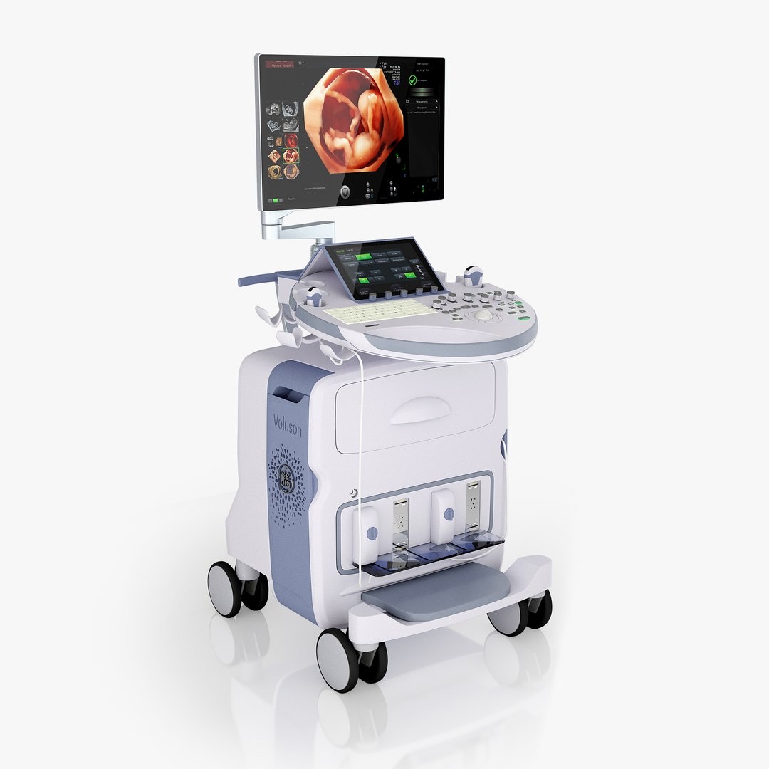
| Ultrasound | GE Voluson E10 with colour Doppler |
|---|
| Preparation | No fasting, no full bladder; bring earlier scan reports |
|---|
| Scan time | Usually about 30-45 minutes, sometimes longer; more if combined with another scan |
|---|
| Radiation | None - ultrasound uses sound waves |
|---|
| Report | Most reports the same day, sent to your WhatsApp |
|---|
| Price | Confirmed on WhatsApp before your visit |
|---|
| Hours | Mon-Sat 8am-10pm, Sun 8am-5pm |
|---|
| Access | Ground floor, step-free |
|---|
What a fetal echo checks
A fetal echo looks only at your baby's heart, step by step, and uses colour Doppler to show which way the blood flows. It can pick up many significant heart conditions before birth, such as a larger hole between the pumping chambers (VSD), tetralogy of Fallot, transposition of the great arteries and some rhythm problems.
- The four chambers and the walls between them.
- The four valves: whether they open, close and seal normally.
- The two main arteries (aorta and pulmonary artery), which should cross each other, and the arch of the aorta.
- The veins bringing blood back to the heart.
- Heart rate and rhythm, and how well the heart pumps.
Which week is a fetal echo done?
Most fetal echos are done at around 18-24 weeks, often at about 20-22 weeks. By then the heart is large enough to see in detail; later in pregnancy the baby is bigger and its bones are harder, so some views become more difficult. This timing also leaves time for advice and planning if something is found, which is why the fetal echo is often done on the same day as the anomaly (TIFFA) scan. It can also be done later if a concern appears. Your obstetrician decides the timing, so tell us the week they asked for when you book.
Why your doctor may ask for it
A fetal echo is advised when there is a higher chance of a heart condition, or when another scan shows that the heart needs a closer look. Many babies born with heart conditions have no risk factor, so some obstetricians advise it more widely.
- A parent, brother or sister of the baby with a heart condition present from birth.
- Diabetes that was present before pregnancy or found in the first three months of pregnancy.
- Certain medicines in early pregnancy, such as some epilepsy medicines or vitamin A-based (retinoid) acne medicines.
- Pregnancy through IVF or other assisted conception.
- A high NT measurement or other heart-related findings at the NT scan, or a possible heart finding on the anomaly scan.
- Another finding in the baby that can go with heart conditions, such as fluid build-up in the baby, or a suspected chromosome or genetic condition.
- A heartbeat that is too fast, too slow or irregular.
- Certain antibodies in the mother (anti-Ro), for example with lupus or Sjögren's syndrome, which can affect the baby's heart rhythm.
- Identical twins who share one placenta (monochorionic twins), or other twin pregnancies as your obstetrician advises.
A fetal echo is a planned scan, not an emergency test. If your baby is moving less than usual, or you have bleeding, leaking fluid, severe tummy pain, a severe headache, blurred vision or fits, do not wait for a scan: go straight to the labour ward or a hospital emergency department first, and let your obstetrician know.
How to prepare
No special preparation is needed. Our team confirms the exact preparation when you book on WhatsApp.
- Eat and drink normally. You do not need to fast or come with a full bladder.
- Bring your obstetrician's prescription and all earlier pregnancy scan reports, especially the NT and anomaly scans.
- If a parent or an earlier child has a heart condition, bring their heart reports if you have them.
- Bring a list of your medicines. If you have diabetes, also bring your sugar reports.
- Tell us if you are expecting twins or if the fetal echo is to be combined with another scan, so that enough time is set aside.
What happens during the scan
You lie on the couch and gel is spread on your tummy. The radiologist moves the probe over your bump to view the heart from several angles, then uses colour Doppler to see the blood flow. The scan should not hurt, though you may feel some pressure from the probe. If lying flat on your back makes you feel dizzy or unwell, tell the radiologist. Turning slightly onto your side usually helps.
It usually takes about 30-45 minutes, and longer when combined with an anomaly or growth scan. If the baby is lying with its back towards your tummy, you may be asked to walk around and come back. Occasionally a repeat scan after a week or two is suggested. You are welcome to bring your partner or a family member.
What a fetal echo cannot show
A normal fetal echo is reassuring, but no scan can detect every heart problem before birth. A fetal echo looks at the heart only: it does not replace the anomaly scan for the rest of the baby, and it does not test the baby's chromosomes or genes. After birth, get your newborn checked by a doctor straight away if they have breathing difficulty, bluish lips or skin, poor feeding, or sweating or tiring during feeds, even if the fetal echo was normal.
- Small holes in the heart are often not visible, especially between the upper chambers, where an opening is normal before birth.
- The ductus, a vessel between the aorta and pulmonary artery, is normally open before birth, so whether it closes properly can only be checked after birth.
- Some problems, such as some valve narrowings, develop or get worse later in pregnancy. Narrowing of the aorta (coarctation) can be hard to confirm until after birth.
- The baby's position, the mother's body build, tummy surgery scars, low fluid or twins can limit the views.
If a heart problem is found
The radiologist explains what was seen before you leave, in English, Hindi or Marathi, and you share the report with your obstetrician. Not every finding is serious, and some small holes close on their own. If a significant condition is suspected, your obstetrician will usually refer you to a fetal medicine specialist and a paediatric cardiologist (a children's heart specialist) to confirm the diagnosis and explain what it means. Because some heart conditions are linked with genetic conditions, your obstetrician may also discuss further tests with you. If needed, the birth can be planned at a hospital with a newborn heart team, and many heart conditions found before birth can be treated after the baby is born.
Is it safe?
Yes. Ultrasound uses sound waves, not radiation, and no injection or dye is used. Colour Doppler is used only for a medical reason, at the lowest setting and for the shortest time that still gives a reliable result, following international ultrasound safety guidance. Used this way, it has no known harmful effect on the baby.
Fetal echo at AV Scan
- Performed and reported by our FMF (UK) certified radiologists: Dr. Avinash Bansal, whose work includes fetal medicine and Doppler, and Dr. Vaishnavi Bansal, whose work includes pregnancy scans.
- Scanned on the GE Voluson E10 ultrasound. It can be done in the same visit as your anomaly (TIFFA) or growth scan.
- Most reports the same day, sent to your WhatsApp, so you can share them with your obstetrician.
- The exact price is confirmed on WhatsApp before your visit. Cashless with most major insurers and TPAs; empanelled for CGHS, ECHS and MJPJAY.
- Open Monday to Saturday 8am-10pm and Sunday 8am-5pm.
- Ground floor and step-free, near Shivaji Park and Siddhivinayak Temple.