At a Glance
| Also called | MRA brain, MRI brain with angiography, MR angio; MRV or MR venography for the veins |
|---|
| What it shows | Brain arteries (MRA) and veins (MRV): aneurysm, narrowing, AVM, clot in the veins |
|---|
| Injection | Usually none for MRA; contrast is sometimes used for MRV or for a closer look |
|---|
| Radiation | None |
|---|
| Time | Usually about 30-45 minutes with a brain MRI; a little longer if MRV or contrast is added |
|---|
| Preparation | No fasting for a plain study; our team confirms the exact preparation on WhatsApp |
|---|
| Reported by | Dr. Avinash Bansal, MD (Radiology) |
|---|
| Report | Most reports the same day, sent to your WhatsApp |
|---|
| Price | Confirmed on WhatsApp before your visit |
|---|
| Payment | Cashless with most major insurers/TPAs; empanelled for CGHS, ECHS, MJPJAY |
|---|
| Hours | Mon-Sat 8am-10pm, Sun 8am-5pm |
|---|
What MRA and MRV of the brain show
MRA shows the arteries that carry blood to the brain, including the ring of connecting arteries at its base called the circle of Willis. It usually uses a time-of-flight (TOF) technique, which builds the picture from the movement of flowing blood, so no dye is normally needed. MRV shows the veins and large venous channels (sinuses) that drain blood from the brain.
Prescriptions often say "MRI brain with angiography" or "MRI brain with MRA/MRV", so the brain tissue and its blood vessels are checked in one sitting. The scan can show:
- Aneurysm: a small balloon-like bulge in the wall of a brain artery
- Narrowing or blockage of arteries inside the head
- Arteriovenous malformation (AVM): an abnormal tangle of arteries and veins
- A clot or narrowing in the brain's veins and venous sinuses (on MRV)
- Normal variations in how the arteries are arranged, which are common and usually harmless
When doctors ask for an MRI brain with angiography
Your neurologist, neurosurgeon or physician may request it:
- To look for an aneurysm when symptoms or an earlier scan raise the question, or to follow up a known one, including after coiling
- For screening when two or more first-degree relatives (parents, brothers, sisters or children) have had a brain aneurysm, including one that has bled (subarachnoid haemorrhage), and in some adults with polycystic kidney disease, particularly with such a family history
- After a stroke or TIA (mini-stroke), to check the arteries inside the head
- When a brain MRI or CT has shown a bleed or a possible abnormal vessel
- Suspected clot in the brain's veins (MRV), for example a persistent, worsening headache with blurred vision, particularly after childbirth, while taking hormone tablets (including the contraceptive pill) or with a clotting disorder
- Raised pressure inside the head (idiopathic intracranial hypertension), to rule out a clot or narrowing in the veins, or a pulsing sound in one ear in time with the heartbeat (pulsatile tinnitus)
- Trigeminal neuralgia or hemifacial spasm, together with detailed thin-slice pictures of the nerves, to look for a blood vessel pressing on a nerve
Emergency first: a sudden severe headache ("the worst headache of my life"), sudden weakness or numbness of the face, arm or leg, difficulty speaking, sudden loss of vision, confusion or drowsiness, a first seizure, a severe new headache during pregnancy or in the weeks after delivery, or a severe headache after a head injury can mean a stroke, a bleed or a clot. Please go to a hospital emergency department straight away, not to an outpatient scan.
How to prepare
For a plain MRI brain with MRA there is no special preparation: eat normally and take your usual medicines. Our team confirms the exact preparation for your scan when you book on WhatsApp.
- If contrast may be needed (more often for MRV), you may be asked not to eat for about 4 hours beforehand; you can usually still drink water and take your regular medicines. A recent kidney function (serum creatinine) report may be requested.
- Tell us before booking about a pacemaker or defibrillator, cochlear implant, aneurysm clip, programmable shunt, nerve stimulator, insulin pump or glucose sensor, any other implanted device, or metal fragments in the eye (for example from welding or grinding work) or elsewhere in the body. Bring the implant card or operation notes.
- Tell us if you are or might be pregnant, are breastfeeding, have kidney disease or allergies, have reacted to contrast before, or take diabetes medicines such as metformin.
- Bring your prescription and earlier MRI, MRA or CT reports and images, especially when an aneurysm is being followed up.
- Wear loose clothes without metal. Jewellery, hairpins, hearing aids and removable dentures with metal parts come off before the scan; some medicine skin patches have a metal layer and may need to come off too, so ask your doctor and bring a spare.
What happens during the scan
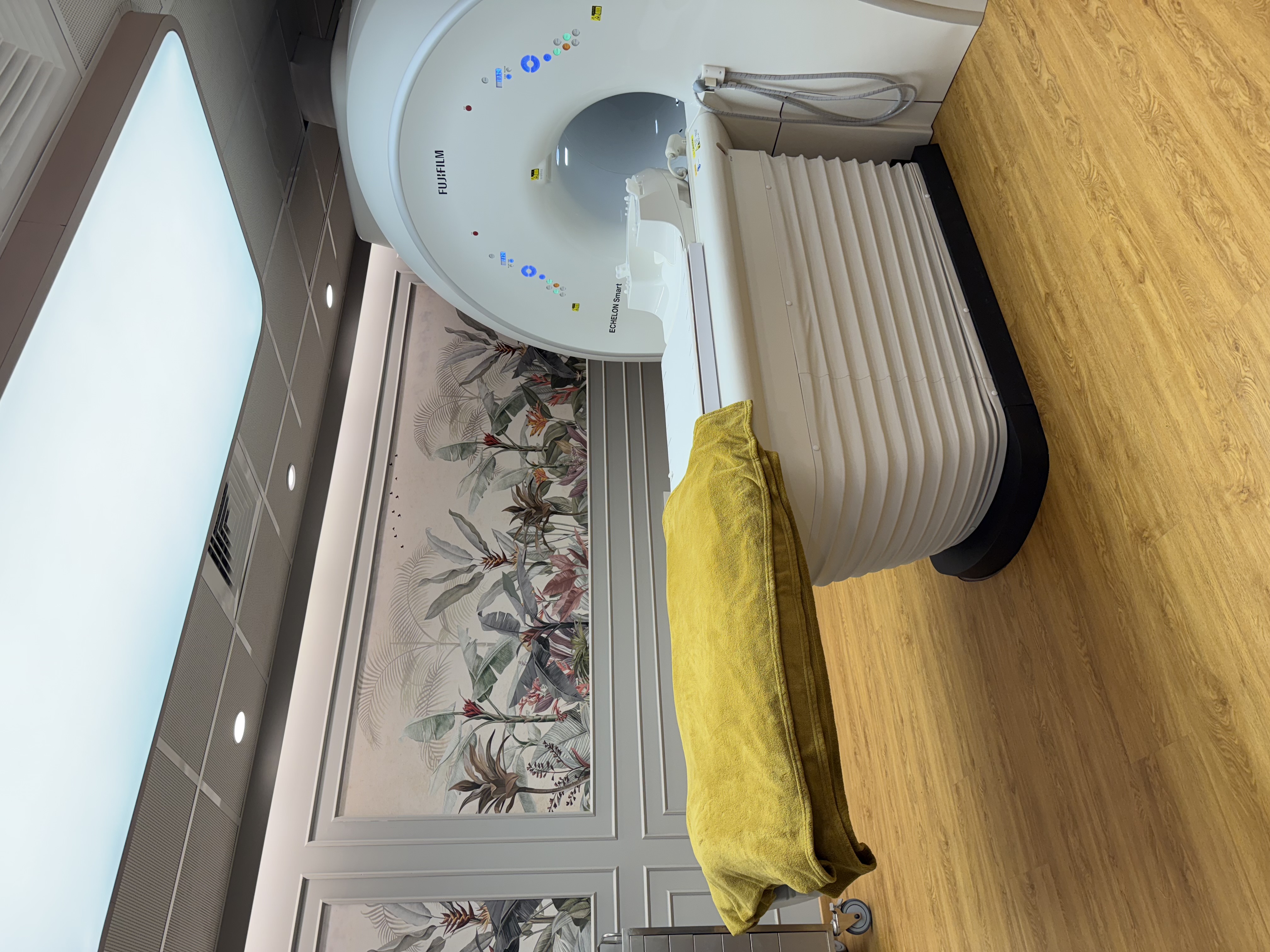
After a short safety check you change into a gown and lie on your back with a lightweight head coil around your head. Please keep your head as still as you can, as movement blurs the vessel pictures. The Fujifilm ECHELON Smart 1.5T uses quieter scanning and you can listen to music; you hold a call bell, and the MRI technologist can see and talk to you throughout.
A brain MRI with MRA usually takes about 30-45 minutes; adding MRV or contrast takes a little longer. Contrast, if needed, is given through a small cannula in an arm vein. Afterwards you can usually go home, eat normally and carry on with your day. If you took a sedative tablet for anxiety, an adult should take you home and you should not drive that day.
Contrast and MRI safety
MRI uses a magnetic field and radio waves, not X-rays. Gadolinium contrast may be used for some MRV studies, or when the plain images show something that needs a closer look, such as a suspected tumour or abnormal vessel; you are told before any injection. It is well tolerated by most people, and serious allergic reactions are rare. A recent creatinine report may be requested, especially if you are older or have kidney disease, diabetes or high blood pressure. If you have severe kidney disease or are on dialysis, tell us, as contrast is then used only after careful discussion with your doctor. Metformin does not normally need to be stopped for MRI contrast, but please mention it.
MRI without contrast can be done in pregnancy when your doctor feels it is needed; contrast is generally avoided in pregnancy unless it is essential. If you are breastfeeding, you do not normally need to stop after MRI contrast.
Implants are checked before you enter the MRI room. Aneurysm clips vary and some older ones are not MRI-safe, so we need the clip details from your operation notes first; the clip can also hide the artery right next to it. Aneurysm coils and stents are usually MRI-compatible, but they can blur the pictures in and around them, so a coiled or stented vessel sometimes needs contrast or another test to assess fully. A programmable shunt may need its setting checked by your neurosurgeon after the scan, so arrange this before booking.
MRA, CT angiography or catheter angiography?
All three show the brain's blood vessels. Your doctor chooses based on the clinical question and how urgent it is.
MRA has limits: it can make a narrowing look tighter than it really is, very small vessels and aneurysms may not be clearly seen, and movement blurs the pictures. If the result is uncertain, your doctor may suggest one of the other tests.
- MRA: no radiation and usually no injection, which suits screening and repeat follow-up, but it takes longer and needs you to lie still.
- CT angiography: very quick, but uses X-rays and iodine contrast; often used in emergencies.
- Catheter angiography (DSA): a thin tube is passed into the arteries in hospital. It is the reference test but carries a small risk, so it is usually kept for planning treatment or when other scans leave a doubt.
MR angiography at AV Scan, Dadar West
Brain MRA and MRV studies are reported by Dr. Avinash Bansal (MD Radiology), who reviews the vessels in 3D reconstructions from several angles alongside the brain images, and compares them with any earlier scans you bring. For the brain-tissue part of the scan (stroke, tumours, epilepsy, headache), see our brain MRI page.
- Fujifilm ECHELON Smart 1.5T MRI, with quieter scanning and in-scanner music
- The exact price is confirmed on WhatsApp before your visit, including whether MRV or contrast is part of your doctor's request
- Most reports the same day, sent to your WhatsApp
- Cashless with most major insurers and TPAs, and empanelled for CGHS, ECHS and MJPJAY: tell us your insurer or scheme on WhatsApp and we will confirm what applies
- Open Monday to Saturday 8am-10pm and Sunday 8am-5pm
- Ground floor and step-free, on Shankar Ghanekar Marg near Shivaji Park and Siddhivinayak Temple