At a Glance
| Also called | USG pelvis, pelvic sonography, TVS, TAS |
|---|
| Reported by | Dr. Vaishnavi Bansal, MD Radiology |
|---|
| Radiation, injection or contrast | None for a routine pelvic scan |
|---|
| Preparation | Full bladder (transabdominal) or empty bladder (TVS); confirmed on WhatsApp |
|---|
| Time on the couch | Usually about 15-30 minutes |
|---|
| Report | Most reports the same day, sent to your WhatsApp |
|---|
| Price | Confirmed on WhatsApp before your visit |
|---|
| Insurance | Cashless with most major insurers/TPAs; CGHS, ECHS, MJPJAY |
|---|
Abdominal or transvaginal scan: what happens
Your prescription and symptoms decide which you need, and many women have both in one visit; the radiologist explains before starting. The scan usually takes about 15-30 minutes on the couch, and you can carry on normally afterwards. A routine pelvic ultrasound uses no radiation, injection or contrast and has no known harmful effects.
A transvaginal scan is done only with your consent, and you can ask to stop at any time. If you would like a female staff member in the room, please tell us when you book. If you have never had intercourse, the scan is done through the abdomen instead.
- Transabdominal: you lie on your back while the probe moves over the lower abdomen with gel. A full bladder can feel uncomfortable for this brief part, but it pushes the bowel aside and shows the whole pelvis, including large fibroids or cysts.
- Transvaginal (TVS): you empty your bladder and lie with your knees bent. A slim probe with a fresh protective cover is placed a short way into the vagina. Lying close to the uterus and ovaries, it shows the lining, small fibroids, polyps and follicles in finer detail.
What a pelvic ultrasound shows
In Hindi it is often called बच्चेदानी की सोनोग्राफी, and in Marathi गर्भाशयाची सोनोग्राफी. The report describes:
- Uterus: size, shape and position, including shape differences present from birth
- Fibroids: number, size and position (bulging into the cavity, within the wall or on the outer surface), which helps plan treatment
- Adenomyosis: lining tissue growing into the muscle wall, often with heavy, painful periods
- Endometrium (lining): thickness, texture, polyps and any fluid in the cavity
- Ovaries: simple cysts, blood-filled cysts from the normal cycle, 'chocolate cysts' (endometriomas), dermoid cysts, and features that need a closer look
- The position of a Copper-T or hormonal coil
- Fluid in the pelvis, swollen fluid-filled tubes, and the urinary bladder
- With a 3D scan, if your gynaecologist asks for it: the shape of the uterine cavity and the outline of the uterus
What a routine scan cannot show
Normal fallopian tubes usually cannot be seen, so a routine scan cannot show whether they are open. Endometriosis often does not show on a routine scan, so a normal scan does not rule it out. If the findings are unclear, if fibroids are very large, or before some treatments, your doctor may ask for an MRI pelvis.
PCOS on ultrasound: what the scan can and cannot tell you
PCOS (polycystic ovary syndrome) is diagnosed by your doctor, not by the scan alone. Under current international guidelines, an adult needs two of three features once thyroid, prolactin and other causes are ruled out: irregular or absent periods; signs of raised male-type hormones (excess facial or body hair, acne, or raised blood levels); and polycystic ovaries on ultrasound, for which some doctors use an AMH blood test instead.
The 'cysts' are really many small egg follicles. The report counts them, more reliably on a TVS, and measures each ovary. In the first 8 years after periods begin, many follicles are normal, so the scan is not used to diagnose PCOS then. Tell us if you take the contraceptive pill, as it changes how the ovaries look.
When doctors request a pelvic ultrasound
Common reasons include:
- Heavy, painful or irregular periods, or bleeding between periods
- Pain or a lump low in the abdomen
- Suspected fibroids, adenomyosis or an ovarian cyst, or follow-up of a known one
- Suspected PCOS, or difficulty conceiving
- Any bleeding after menopause, which should always be checked
- Checking a Copper-T or hormonal coil
- Men with urinary symptoms: the bladder, an estimate of prostate size and, if your doctor asks, the urine left after passing urine. The scan cannot show whether prostate cancer is present.
Go to a hospital emergency department first if you have sudden severe pain low in the abdomen (especially on one side, with vomiting), heavy bleeding with dizziness or fainting, lower abdominal pain with fever, or pain or bleeding with a missed period or a positive pregnancy test. These can be signs of a twisted ovary, a burst cyst, a pelvic infection or an ectopic pregnancy, which need urgent care.
How to prepare
Our team confirms the exact preparation when you book on WhatsApp. As a general guide:
- Transabdominal (women and men): drink about 1 litre of water, finishing about an hour before the scan, and do not pass urine until after it. If you have kidney or heart disease or have been told to limit fluids, ask us first.
- Transvaginal: an empty bladder; please remove any tampon or menstrual cup beforehand. If both scans are planned, the abdominal part is done first.
- Abdomen and pelvis together: also avoid food for about 4-6 hours before (plain water is fine). If you have diabetes, ask us how to manage food and medicines on the day.
- Cycle day: follow any day your gynaecologist gives. Otherwise, the lining, fibroids and polyps are usually checked a few days after bleeding stops (around day 5-10), PCOS features around day 2-5, and a cyst recheck when your doctor advises; pain, bleeding or scans after menopause can be any day.
- Wear a two-piece outfit (such as a kurta with trousers), bring previous scan reports, and note the first day of your last period.
- Tell us if you are, or might be, pregnant, have a Copper-T or coil, or have a latex allergy.
Related tests at AV Scan
Other tests often linked to a pelvic scan:
- Pregnancy: if your period is late or your pregnancy test is positive, book an early pregnancy scan instead. Later scans are on our pregnancy scans page.
- Follicle study: short scans across one cycle to track how egg follicles grow and whether ovulation happens. Your doctor decides when it starts.
- Sono-HSG: sterile fluid passed through a thin, soft tube into the uterus outlines the cavity during an ultrasound, and a special bubble-containing fluid shows whether the fallopian tubes are open. It is done only when there is no chance of pregnancy, usually in the days after a period ends, and not during a pelvic infection. Brief period-like cramps are common.
- Kidneys, liver and gallbladder: abdominal ultrasound, often combined with the pelvis in one scan.
- Blood samples for tests your doctor asks for can be given at the centre (see blood tests).
Your pelvic ultrasound at AV Scan, Dadar West
To book, send a photo of your prescription on WhatsApp +91 98200 51511, call the same number, or use our booking page.
- Reported by Dr. Vaishnavi Bansal, MD Radiology, FMF (UK) certified, who looks after breast, women's and fetal imaging
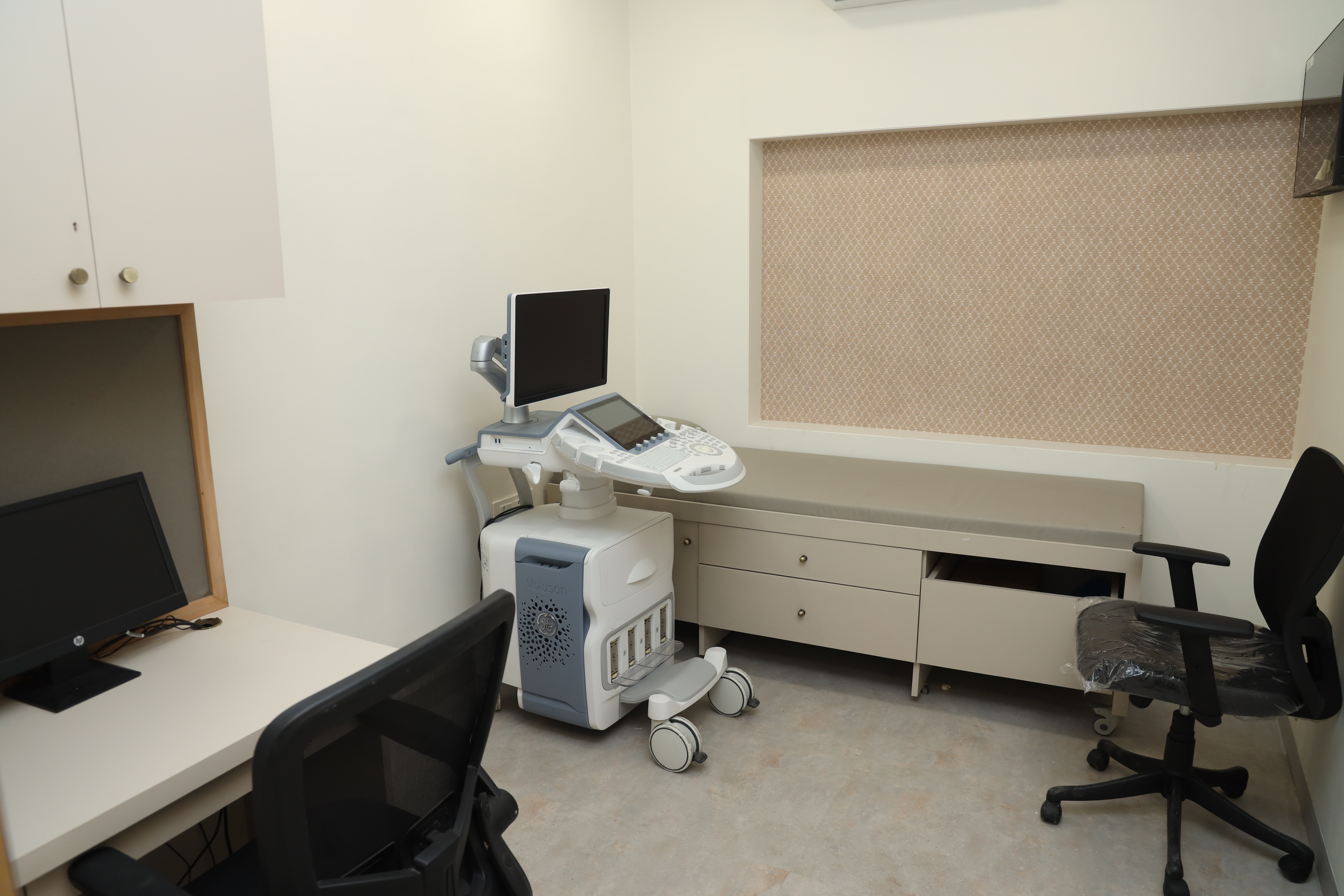
- Ultrasound systems at the centre: GE Voluson E10 and Philips Affiniti 70G
- The exact price is confirmed on WhatsApp before your visit
- Cashless with most major insurers and TPAs; empanelled for CGHS, ECHS and MJPJAY
- Open Monday to Saturday 8am-10pm and Sunday 8am-5pm; most reports the same day, sent to your WhatsApp
- Ground floor, step-free: Shilpa Apartment, Shankar Ghanekar Marg, Dadar West, Mumbai 400028, near Shivaji Park and Siddhivinayak Temple