At a Glance
| Also called | Heart MRI, cardiac magnetic resonance (CMR), MRI heart with contrast |
|---|
| Radiation | None (MRI uses a magnet and radio waves) |
|---|
| Contrast | Usually a gadolinium injection; a recent creatinine report may be requested (the test can be done at the centre) |
|---|
| Time in the scanner | Usually about 45-60 minutes, with ECG stickers and many short breath-holds |
|---|
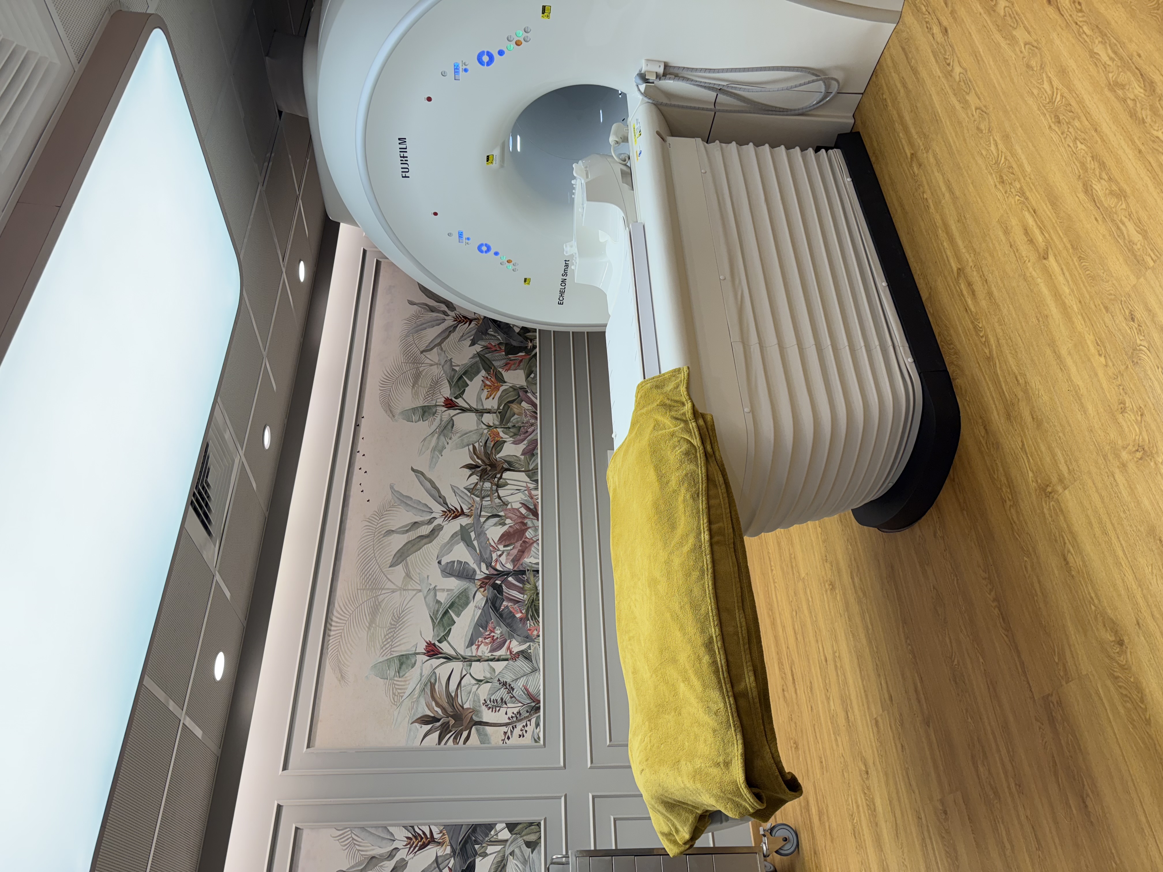
| Scanner | Fujifilm ECHELON Smart 1.5T MRI, quieter scanning, in-scanner music |
|---|
| Reported by | Our radiologists, Dr. Avinash Bansal and Dr. Vaishnavi Bansal |
|---|
| Preparation | Our team confirms the exact preparation when you book on WhatsApp |
|---|
| Report | Most reports the same day, sent to your WhatsApp |
|---|
| Price | Exact price confirmed on WhatsApp before your visit |
|---|
| Payment | Cashless with most major insurers and TPAs; empanelled for CGHS, ECHS and MJPJAY |
|---|
| Hours and access | Mon-Sat 8am-10pm, Sun 8am-5pm; ground floor, step-free |
|---|
What a cardiac MRI shows
Cardiac MRI uses a magnet and radio waves, not X-rays, and times the pictures to your heartbeat, so they play back as short moving films of the heart pumping. Patients also call it a heart MRI (हृदय का MRI स्कैन / हृदयाचा MRI स्कॅन). It can show:
- Pumping function: the size of each pumping chamber, the thickness of the heart muscle and the ejection fraction (the share of blood pumped out with each beat), including the right side of the heart, which echo can find hard to see
- Scarring, using late gadolinium enhancement (LGE): pictures taken several minutes after the contrast injection, in which scarred or damaged muscle shows up bright
- Myocarditis (inflammation of the heart muscle), for example after a viral illness: swelling and patches of damage in the muscle
- Cardiomyopathy (heart muscle disease), such as thickened muscle (hypertrophic) or an enlarged, weak heart (dilated); the pattern of scarring helps tell the types apart
- Damage after a heart attack: where the scar is and how much of the muscle wall it involves, which helps your cardiologist plan treatment
- Heart conditions present from birth (congenital heart disease), usually as follow-up after surgery
When cardiologists request a cardiac MRI
Common reasons your cardiologist may request one:
- An echo showing a weak or enlarged heart, or thickened muscle, where the cause is not clear
- Suspected myocarditis: chest pain, breathlessness or palpitations after a viral infection
- Chest pain with a raised troponin (a blood test for heart muscle damage) when angiography found the heart arteries open
- Palpitations, fainting or an abnormal ECG, to look for a heart muscle cause
- A family history of cardiomyopathy or sudden death at a young age, when your cardiologist advises checking
- Follow-up of a known cardiomyopathy or a heart condition present from birth
Cardiac MRI is a planned test, not an emergency test. If you have chest pain now, chest pain with sweating or breathlessness, sudden severe breathlessness, fainting, or a racing heartbeat with dizziness, go to the nearest hospital emergency department first. Do not wait for a scan appointment.
Cardiac MRI, 2D echo and other heart tests
A 2D echo is an ultrasound of the heart: quick, with no injection, and the usual first test for pumping strength and the heart valves. Its view depends on a clear window between the ribs and lungs, and it cannot show scar in the muscle the way MRI can. The two are used together; MRI does not replace echo.
CT coronary angiography, a type of CT angiography, uses X-rays and an iodine contrast injection to look for narrowing in the heart arteries, and a TMT (treadmill test) records your ECG during exercise. Cardiac MRI looks mainly at the heart muscle and is not the usual test for blocked arteries; your cardiologist chooses the test that answers their question.
How to prepare for a cardiac MRI
Our team confirms the exact preparation when you book on WhatsApp. As a general guide:
- Food: our team tells you whether to avoid food for a few hours beforehand. Water is usually fine.
- Take heart and blood pressure medicines as usual unless your cardiologist has said otherwise; do not stop any medicine on your own. Tell us if you take insulin or diabetes tablets.
- Bring your prescription, echo and ECG reports, any earlier heart scan or angiography reports, discharge summaries, a list of your medicines and any implant or heart device card.
- Wear loose clothing without metal; you will change into a gown and remove jewellery, watches and hearing aids. Tell us about any medicine skin patch, insulin pump or glucose sensor: some contain metal and must be taken off, so ask your doctor beforehand how to manage this.
- Tell us if your heartbeat is irregular or very fast, if lying flat or holding your breath is difficult, or if closed spaces make you anxious.
What happens during the scan
After a metal-safety checklist, a small cannula is placed in an arm vein. ECG stickers on your chest let the scanner time each picture to your heartbeat; a small patch of chest hair may need trimming. You lie on your back with a light receiver pad (coil) over your chest, ear protection on and a call bell in your hand.
You will be asked to hold your breath many times, usually for about 10-15 seconds each, because breathing blurs the pictures; an irregular or very fast heartbeat can also blur them. Our team adjusts the scan to reduce this, but picture quality can vary. Contrast is given partway through, with the scar pictures several minutes later. The scan usually takes about 45-60 minutes in the scanner, with music to listen to. Afterwards you can usually eat, drive and carry on as normal; after sedation, you will need an adult to take you home and should not drive for the rest of the day.
Contrast and MRI safety
Gadolinium, the MRI contrast, is different from the iodine dye used for CT; reactions are uncommon and usually mild, and our team is prepared to manage them. A recent kidney function (serum creatinine) report may be requested; if you do not have one, the test can be done at the centre before your scan. Tell us about any allergy or previous contrast reaction, asthma, kidney disease or dialysis (in severe kidney disease our radiologists weigh the need for contrast carefully), and diabetes medicines such as metformin: we check your kidney function (eGFR) and advise you. Tell us if you are, or might be, pregnant, as gadolinium is generally avoided in pregnancy unless essential. If you are breastfeeding, tell us; current guidance says you can usually continue breastfeeding normally after gadolinium.
Pacemakers and defibrillators (ICDs): tell us before booking and bring your device card. Some MR-conditional devices can be scanned under supervision, following the device's conditions; others cannot. A device that can be scanned usually has to be switched to an MRI setting before the scan and back afterwards, so we need its details well in advance; it can also blur parts of the heart pictures, so your cardiologist may prefer a different test. Coronary stents, most replacement heart valves and breastbone wires from heart surgery are generally not a problem, but tell us about them, and about any loop recorder, cochlear implant, nerve stimulator, aneurysm clip or metal in the eye.
If closed spaces make you anxious, sedation is available when needed, under the supervision of an anaesthesiologist; because cardiac MRI relies on breath-holds, our radiologists will advise whether it suits your scan.
Cardiac MRI at AV Scan, Dadar West
Share your cardiologist's prescription on WhatsApp, call +91 98200 51511 or use our booking page; our radiologists plan the scan around the question on it. Our team confirms the preparation, whether contrast is planned, and the exact price before your visit.
- Reported by our radiologists, Dr. Avinash Bansal and Dr. Vaishnavi Bansal, both MD (Radiology) and FMF (UK) certified
- Fujifilm ECHELON Smart 1.5T MRI, with quieter scanning and in-scanner music
- 2D echo, ECG, TMT, CT angiography and creatinine blood tests at the same centre
- Cashless with most major insurers and TPAs; empanelled for CGHS, ECHS and MJPJAY
- Most reports the same day, sent to your WhatsApp
- Open Monday to Saturday 8am-10pm and Sunday 8am-5pm
- Ground floor, step-free: Shilpa Apartment, Shankar Ghanekar Marg, Dadar West / Prabhadevi, near Shivaji Park and Siddhivinayak Temple