At a Glance
| Most new back pain | Usually settles in a few weeks; an early scan rarely changes treatment |
|---|
| X-ray | Bones: alignment, wear and tear, fractures, a slipped vertebra. Does not show discs or nerves |
|---|
| MRI | Discs, nerves, spinal cord and bone marrow: the test for sciatica and a slipped disc. No radiation |
|---|
| CT | Fine bone detail, or when MRI is not possible. More radiation than an X-ray |
|---|
| Go to emergency first | Loss of bladder or bowel control, numbness around the private parts, new leg weakness, fever with back pain, major injury, sudden severe pain with feeling faint |
|---|
| Who decides | Your doctor, after examining you |
|---|
| Reported by | Our radiologists, Dr. Avinash Bansal and Dr. Vaishnavi Bansal |
|---|
| Report | Most reports the same day, sent to your WhatsApp |
|---|
| Price | Exact price confirmed on WhatsApp before your visit |
|---|
| Open | Mon-Sat 8am-10pm, Sun 8am-5pm; ground floor, step-free |
|---|
Do you need a scan for back pain at all?
Most back pain comes from the muscles, ligaments and small joints of the spine, and eases within a few weeks with gentle activity, the painkillers your doctor advises and, often, physiotherapy. Even sciatica, pain travelling from the back down the leg, often improves over several weeks. So unless there are warning signs, doctors usually do not ask for a scan early on: it seldom changes the treatment. Your doctor may still advise a scan sooner if the pain is severe, getting worse or not improving as expected.
Scans also show disc bulges and wear and tear (spondylosis) in many adults with no back pain, more so with age. Your doctor matches the report with your symptoms and examination to decide what is causing the pain.
In Hindi, lower back pain is often called कमर दर्द; in Marathi, कंबरदुखी.
Warning signs: go to a hospital emergency department first
Do not wait for a routine scan appointment if you have back pain with any of these:
- Loss of bladder or bowel control, new difficulty passing urine, or leaking urine
- Numbness around the private parts, back passage or inner thighs
- New or worsening weakness in a leg or foot, such as a foot that drags, new unsteadiness when walking, or sciatica in both legs
- Fever, or feeling very unwell, with back pain
- Back pain after a major injury, such as a road accident or a fall from a height
- New back pain with leg weakness or numbness if you have had cancer
- Sudden severe back or tummy pain with feeling faint
These can mean pressure on the nerves at the base of the spine (cauda equina syndrome) or on the spinal cord, infection, a serious injury, or a problem with the main blood vessel in the abdomen. They need urgent hospital assessment, so go straight to the emergency department; the emergency team arranges any urgent scan. If you have had cancer and develop new back pain, even without these signs, contact your cancer doctor promptly.
When an X-ray helps
An X-ray shows the bones: alignment, wear and tear, a curve (scoliosis), a vertebra slipped forward on the one below (spondylolisthesis), and many fractures, including a vertebra collapsed by thin bones (osteoporosis). It does not show discs, nerves or the spinal cord, so it cannot show a 'slipped disc' or confirm sciatica, and a normal X-ray does not rule out early infection or tumour.
Doctors commonly ask for an LS spine (lower back) X-ray to check alignment, before and after spine surgery, or after a fall in an older person or someone with osteoporosis or on long-term steroid tablets, in whom even a minor fall can cause a fracture. Flexion-extension views, taken as you bend forwards and backwards, check for abnormal movement between the vertebrae. If a vertebra has collapsed, a DEXA scan may follow to measure bone density. The radiation dose of a lower-back X-ray is small but higher than that of a chest X-ray, one reason it is not done routinely for simple back pain; more on our digital X-ray page.
When an MRI is advised
MRI shows the discs, nerve roots, spinal cord, bone marrow and soft tissues, without radiation. It is the test for a slipped disc (disc prolapse), a pinched nerve, narrowing of the spinal canal (stenosis), and infection or tumour. The usual request is a lumbar spine MRI ('MRI LS spine'); doctors add whole-spine screening to check the rest of the spine, for example when more than one area may be involved. Doctors commonly ask for MRI for:
- Leg pain, numbness or tingling that has not settled after about six weeks of treatment, especially if an injection or operation is being considered
- Weakness in the leg or foot (if it is new or getting worse, go to the emergency department first)
- Legs that ache or go numb on walking and ease on sitting or bending forwards, a pattern seen with spinal stenosis
- Suspected infection (including spinal tuberculosis), tumour or spread of cancer: for example back pain with fever, weight loss, pain worse at night, or past cancer
- A collapsed vertebra on X-ray, to see whether it is new or old and why it happened
- Planning or follow-up of spine surgery
When CT is used
CT shows bone in fine detail: complex fractures, a small crack in the back of a vertebra (spondylolysis, seen in some young sportspeople), fusion after surgery and the position of screws. In young people MRI is often tried first for a suspected crack, to avoid radiation, with CT used if the answer is still unclear. CT shows discs less clearly than MRI and nerves poorly, so it is not the first test for sciatica, but it helps when MRI is not possible, for example with some implants. Its radiation dose is higher than an X-ray's; see CT scan radiation explained.
Back pain that may not come from the spine
Pain in the side of the back that comes in waves and spreads towards the groin may be a kidney stone, which needs different tests; see which scan for kidney stones. Pain in the side of the back with fever, shivering or burning urine can be a kidney infection and needs a doctor the same day.
Preparing, safety and contrast
Our team confirms the exact preparation when you book on WhatsApp. Spine X-rays and plain MRI or CT scans usually need no fasting. Take your usual medicines, including painkillers, since lying still is easier when pain is controlled, and bring earlier spine scans and reports.
Tell us before any X-ray or CT if you are or might be pregnant. Before you book an MRI, tell us about a pacemaker, a cochlear implant, a brain aneurysm clip, any metal fragment in the eye, a nerve stimulator, spinal rods or any other implant, and bring the device card: some pacemakers and MR-conditional implants can be scanned here under supervision, following the device's conditions; others cannot. Sedation is available when needed, under the supervision of an anaesthesiologist. See our MRI preparation guide.
Most spine MRIs and CTs need no contrast; for MRI it may be used for suspected infection or tumour, or after surgery to tell scar tissue from a new disc prolapse. If contrast is planned, a recent kidney function (creatinine) report may be requested; if you have none, the test can be done at the centre before the scan. Tell us about kidney disease, asthma, allergies or a past contrast reaction, and your diabetes medicines, especially metformin: for CT contrast we check your kidney function (eGFR) and advise you. A CT with contrast needs about 4 hours without food (water is allowed). MRI contrast is avoided in pregnancy unless essential.
Your back pain scan at AV Scan
Your doctor decides which scan you need. We advise seeing a doctor before booking a spine MRI; X-rays and CT scans use radiation and are done only when a doctor has asked for them. WhatsApp us a photo of the prescription and our team confirms the study, the preparation and the exact price before your visit.
- Reported by our radiologists, Dr. Avinash Bansal and Dr. Vaishnavi Bansal; musculoskeletal (bone, joint and spine) imaging is led by Dr. Avinash Bansal
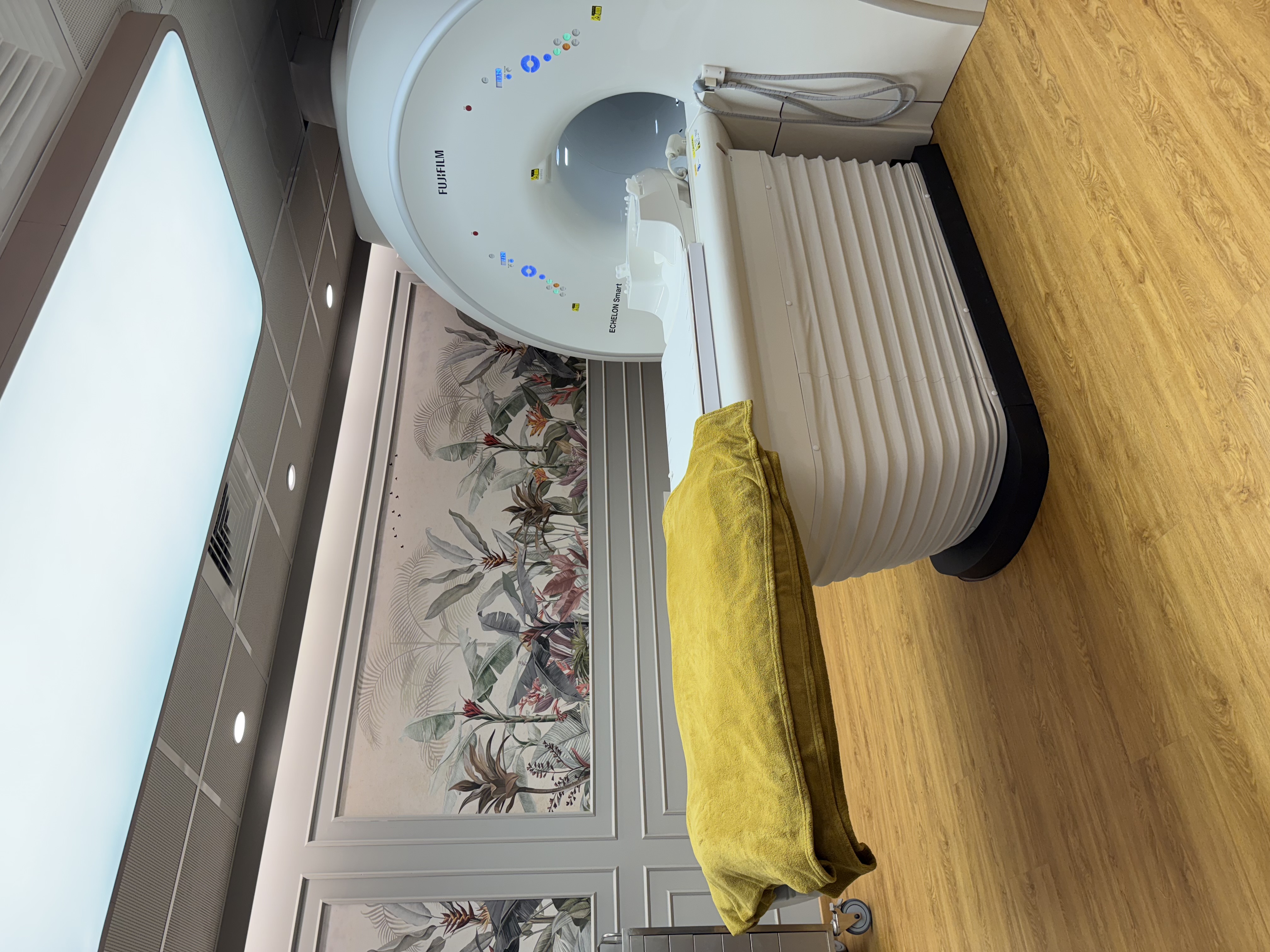
- Fujifilm ECHELON Smart 1.5T MRI with quieter scanning and in-scanner music; Fujifilm Supria CT; digital X-ray, with walk-ins welcome on a prescription
- Most reports the same day, sent to your WhatsApp
- Cashless with most major insurers and TPAs; empanelled for CGHS, ECHS and MJPJAY
- Open Monday to Saturday 8am-10pm and Sunday 8am-5pm
- Ground floor with step-free entry, which helps when your back hurts; near Shivaji Park and Siddhivinayak Temple