
CT-Guided Biopsy & FNAC in Dadar West, Mumbai
A CT-guided biopsy or FNAC takes a small sample from a deep area that ultrasound cannot show clearly, with CT pictures guiding the needle to the right spot. At AV Scan in Dadar West, our radiologists do the procedure, with a local anaesthetic to numb the skin. A pathologist then examines the sample, and your doctor explains the result.
At a glance
- Also called
- CT-guided needle biopsy, CT-guided core biopsy, CT-guided FNAC, percutaneous (through the skin) biopsy
- Done by
- Our radiologists, MD (Radiology)
- Scanner
- Fujifilm Supria CT, with the dose kept as low as reasonably achievable
- Pain relief
- A local anaesthetic numbs the skin; most people feel pressure rather than pain
- Time
- Usually under an hour, plus time for consent and for rest afterwards
- Preparation
- Tell us about any blood thinner when you book; never stop one yourself
- Radiation
- Yes (CT uses X-rays); only the area around the target is scanned
- Result report
- From a pathology laboratory; our team tells you when to expect it
- Clinic hours
- Mon–Sat 8am–10pm, Sun 8am–5pm; your procedure day and time are fixed when you book
- Cost
- We tell you the exact figure on WhatsApp before you book.
What is a CT-guided biopsy or CT-guided FNAC?
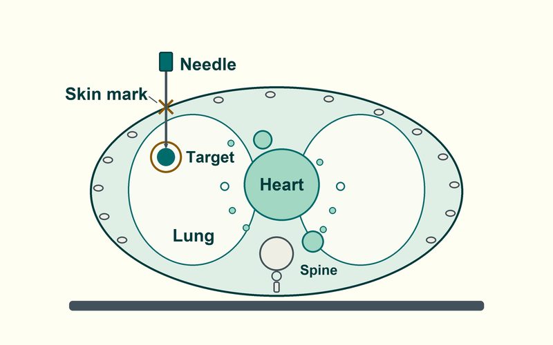
A biopsy takes a small piece of tissue, or a few cells, from an abnormal area for examination under a microscope. In a CT-guided biopsy the radiologist passes a thin needle through the skin, and CT pictures show where the needle tip is at each step [1]. Biopsy is बायोप्सी in both Hindi and Marathi.
- CT-guided FNAC (fine needle aspiration cytology): a very fine needle draws out fluid or clusters of cells.
- CT-guided core biopsy: a slightly larger, spring-loaded needle takes thin cores of tissue, on which the laboratory can run extra tests. In lung cancer, for example, these include marker (biomarker) tests that help choose treatment [1] [9].
A needle biopsy is less invasive than an operation, needs no stitches, and its results are considered as accurate as a surgical sample [1], although sometimes, especially when lymphoma is suspected, more tissue is needed [1].
CT or ultrasound guidance: which one is used?
At AV Scan most needle tests are guided by ultrasound, which shows the needle moving live with no radiation (see ultrasound-guided biopsy and FNAC). Ultrasound cannot see through air or bone, and its picture fades with depth [4], so a small number of deeper targets that ultrasound cannot show clearly are sampled under CT guidance instead:
| Where the target is | Usual guidance |
|---|---|
| Near the surface: a thyroid nodule, a neck or armpit lymph node, a breast lump (our breast imaging specialist advises the right needle test) | Ultrasound |
| In the lung | CT: air blocks ultrasound [4]; a lump touching the chest wall can sometimes be sampled under ultrasound [2] |
| In or behind bone, such as the spine or pelvis | CT: bone blocks ultrasound [4] |
| Deep in the abdomen or pelvis, or behind bowel gas | CT when ultrasound cannot show it clearly [4]; many liver and kidney targets are sampled under ultrasound [12] [1] |
| A small, deep nodule near blood vessels or airways | CT, for careful planning of the needle path [2] |
The radiologist makes the final choice after looking at your scans.
Why doctors ask for a CT-guided biopsy
A scan shows where an abnormal area is; often only a sample can say what it is: harmless, an infection, inflammation or cancer [1]. Common reasons:
- A lung nodule or mass on a chest X-ray or HRCT chest, especially one that a bronchoscopy cannot reach [2]
- A bone lesion, for example in the spine or pelvis, to tell an infection from a tumour [3]
- Enlarged lymph nodes deep in the chest or abdomen, or a deep abdominal mass [1]
- Suspected tuberculosis (TB), a common non-cancer finding in an Indian study of CT-guided FNAC [8]; in India lung cancer is sometimes mistaken for TB, which delays treatment [9]
Whether your target can be sampled at AV Scan depends on where it is and what your scans show; the radiologist reviews them, and our team tells you when you book. Still need the scan itself? See CT scans at AV Scan.
How to prepare for a CT-guided biopsy
- Send a photo of your doctor's prescription and recent scan reports on WhatsApp (opens WhatsApp); our team confirms the procedure, the preparation and the day.
- Tell us about any medicine that thins the blood or prevents clots, and do not stop it yourself. The doctor who prescribed it weighs the bleeding risk against your risk of a clot and decides whether and when to pause it; deep biopsies generally carry more bleeding risk than surface ones [6].
- Our team may ask for a recent clotting test (PT/INR) and a platelet count; the blood sample can be given at the centre (see blood tests).
- Tell us about a bleeding problem or liver disease, an allergy to local anaesthetic, antiseptic or latex, severe lung disease such as emphysema, or a possible pregnancy [2].
- If contrast is needed, a recent kidney function (creatinine) report may be requested; tell us about any allergy and about any diabetes medicines, such as metformin.
- Bring earlier scans (CD, DVD or cloud link), their reports and any earlier FNAC or biopsy reports. If your CT was done here, the radiologist already has your images to plan the needle route.
- Our team tells you whether you need to avoid food for a few hours beforehand.
- Plan for an adult to take you home, and do not drive yourself [7]; our team tells you whether an escort is essential.
Before you come
Our team confirms the exact preparation for your procedure when you book on WhatsApp.
What happens during a CT-guided biopsy
Consent
The radiologist goes through your scans with you, explains the procedure, its risks and the other options, and answers your questions before you sign a consent form.
Positioning and a planning scan
You lie on the CT table on your back, front or side. A short scan confirms the target and a needle route that avoids blood vessels, airways and bowel, and the entry point is marked on the skin [2] [7].
Numbing the skin
The skin is cleaned and numbed with a local anaesthetic injection, which stings briefly. A tiny nick lets the needle in; no stitches are needed [1].
Guiding the needle and taking samples
The needle moves forward in small steps, with a quick CT check after each. For a chest biopsy, breathe gently, do not cough, and hold your breath the same way each time you are asked. Usually two or three samples are taken, sometimes more [7] [2]; you feel pressure, and a core needle makes a click as each sample is taken.
Rest and checks
A small dressing goes on and you rest while the team checks your breathing and pulse. After a lung biopsy a check scan or chest X-ray is often done before you go home [2] [7]; our team tells you how long to plan for. If there is an urgent finding, the radiologist contacts your doctor directly.
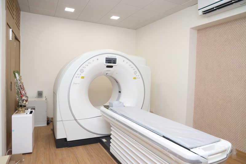
The procedure usually takes under an hour [2]. Say straight away if you feel faint, breathless or in pain.
Risks of a CT-guided biopsy, in plain numbers
Any CT-guided biopsy
- Soreness and a small bruise for a day or two: common.
- Bleeding: usually small. At the skin it stops with pressure, and a little bleeding inside the body usually settles on its own; this is why clotting is checked beforehand [1] [7].
- Infection needing antibiotics: less than 1 in 1,000 [1].
- Injury to a nearby structure, such as the bowel, a nerve or a blood vessel: rare [1] [3].
- Spread of a tumour along the needle track: rare [10].
Extra risks of a CT-guided lung biopsy
The needle crosses the thin lining around the lung, so the main risk is an air leak (pneumothorax, a partly collapsed lung). A review of many studies [5] and an NHS patient leaflet [7] give these figures:
- Air leak (pneumothorax)
- About 1 in 4 people after a core biopsy and 1 in 5 after an FNAC [5]; most are small and settle on their own [7].
- Air leak that needs treatment
- About 1 in 20 people [5] [7]. Treatment may be a small chest tube that lets the air out; it is generally removed the next day [2].
- Coughing up a little blood
- About 1 in 25 after a core biopsy and fewer than 1 in 50 after an FNAC [5]; it fades over 12 to 48 hours [2].
- Bleeding inside the chest that needs an operation
- Very rare: about 1 in 1,000 [7].
- Air entering a blood vessel (air embolism)
- Very rare: about 1 in 3,000 lung biopsies. It can cause chest pain, a heart attack or a stroke [7].
- Death
- Extremely uncommon: about 1 in 5,000 [7].
A core biopsy has more complications than an FNAC, mostly minor, but gives more tissue for tests; the radiologist weighs this for you [5]. A lung biopsy may not suit people with severe emphysema, lung cysts, a clotting disorder, low blood oxygen, high blood pressure in the lungs (pulmonary hypertension) or some kinds of heart failure [2].
Radiation and pregnancy
CT uses X-rays, but only the area around the target is scanned, with the dose kept as low as reasonably achievable on our Fujifilm Supria CT; the small risk is weighed against the benefit of a diagnosis [2]. Tell us if you are or might be pregnant: CT-guided procedures are generally avoided in pregnancy [1].
After your biopsy: aftercare and warning signs
- Keep the dressing dry until the next day [2].
- Rest for the remainder of the day. Avoid heavy lifting and strenuous exercise that night and for one full day after; most people return to normal activities on the second day after the biopsy [2].
- After a lung biopsy, have an adult stay with you overnight [7].
- After a lung biopsy, ask the radiologist before you book a flight; some hospitals advise not flying for up to six weeks [7] [2].
- Restart a blood thinner only when the doctor who prescribed it says so [6].
Contact your doctor or call us for bleeding that does not stop with firm pressure, pain, swelling or redness that keeps increasing, pus at the site, or a fever [3].
Please note
After a chest or lung biopsy, go to the nearest hospital emergency department straight away, or call 108 or 112, if you become more breathless, have sharp chest or shoulder pain when you breathe in, a racing pulse or bluish lips, or cough up more than a little blood [2] [7]. Let us know afterwards.
Your biopsy result and who explains it
The samples go to our partner pathology laboratory, where a pathologist issues a cytology report (on cells, for an FNAC) or a histopathology report (on tissue, for a core biopsy); when TB is possible, part of the sample may be tested for TB germs [8]. This takes longer than a scan report, and special tests such as immunohistochemistry (IHC: special stains that show what type of cells they are) add time. Our team tells you when to expect your result.
The report goes to your referring doctor, who explains it together with your scans.
Sometimes a sample is non-diagnostic: it has too few cells or too little tissue for an answer. That is not a bad sign in itself, and your doctor may advise a repeat biopsy, another test or a surgical biopsy [1]. In two Indian studies of CT-guided FNAC of lung masses, a specific diagnosis was reached in about 86 of 100 patients [8], and about 95 of 100 results matched later tissue tests [11].
Questions about cost?
We tell you the exact figure on WhatsApp before you book. Send a photo of your prescription on WhatsApp (opens WhatsApp), and our team confirms the procedure and the preparation at the same time. See how our quotes work.
Frequently asked questions
What is a CT biopsy test, and how is it different from a CT-guided FNAC?
Both are needle tests under CT guidance. An FNAC draws out cells with a very fine needle; a core biopsy takes thin strips of tissue, on which more laboratory tests can be run. The radiologist chooses the one that suits your target [1].
Is a CT-guided biopsy painful?
The local anaesthetic stings for a moment; after that most people feel pressure rather than pain [2]. Say so if it hurts. Mild soreness for a day or two is common.
How likely is a collapsed lung after a CT-guided lung biopsy?
An air leak around the lung (pneumothorax) happens in about 1 in 4 to 1 in 5 people, and most settle on their own. About 1 in 20 need treatment, such as a small chest tube that is generally removed the next day [5] [2]. Serious complications, such as air entering a blood vessel, are very rare: about 1 in 3,000 [7].
Should I stop my blood thinner before a CT-guided biopsy?
Not on your own. Tell us when you book: the doctor who prescribed it decides whether a short pause is safe for you, because stopping some of these medicines can lead to a clot [6]. Our team may ask for a clotting test.
When can I go back to work, exercise or fly after a CT-guided biopsy?
What happens if the biopsy sample is not enough for a diagnosis?
Where can I have a CT-guided biopsy test in Mumbai?
At AV Scan Diagnostics in Dadar West / Prabhadevi, near Siddhivinayak Temple and Shivaji Park, on a doctor's prescription. Send a photo of the prescription and your scan reports on WhatsApp (opens WhatsApp); our team confirms whether your procedure can be done here, the preparation and the day.
Sources
- Biopsy overview (general biopsy). RadiologyInfo.org (ACR and RSNA).
- Lung biopsy. RadiologyInfo.org (ACR and RSNA).
- Bone biopsy. RadiologyInfo.org (ACR and RSNA).
- General ultrasound. RadiologyInfo.org (ACR and RSNA).
- Complication rates of CT-guided transthoracic lung biopsy: meta-analysis. Heerink WJ et al., European Radiology 27:138-148, doi 10.1007/s00330-016-4357-8, 2017.
- Society of Interventional Radiology consensus guidelines for the periprocedural management of thrombotic and bleeding risk in patients undergoing percutaneous image-guided interventions, Part II. Patel IJ et al., Journal of Vascular and Interventional Radiology 30:1168-1184, 2019.
- CT guided lung biopsy (patient information leaflet 46-2286-V2). NHS patient information, West Hertfordshire, 2024.
- CT guided fine needle aspiration cytology of mass lesions of lung: our experience. Gangopadhyay M et al., Indian Journal of Medical and Paediatric Oncology 32:192-196, 2011.
- Expert consensus on diagnosis and molecular testing strategies for non-small cell lung cancer in India. Biswas B et al., Indian Journal of Medical and Paediatric Oncology 47(2):85-93, doi 10.1055/s-0045-1809375, 2026.
- Tumour seeding following percutaneous needle biopsy: the real story!. Robertson EG, Baxter G, Clinical Radiology 66:1007-1014, 2011.
- Computed tomogram guided fine-needle aspiration cytology of lung mass with histological correlation: a study in Eastern India. Mondal SK et al., South Asian Journal of Cancer 2:14-18, 2013.
- Ultrasound-guided percutaneous liver biopsy: a review of what operators need to know. Alturkistani H et al., Medicine (Baltimore) 103(30):e38673, doi 10.1097/MD.0000000000038673, 2024.
Related tests
Page updated
Book or ask a question
Visit us
AV Scan Diagnostics
Ground Floor, Shilpa Apartment, Shankar Ghanekar Marg, near Shivaji Park, Dadar West / Prabhadevi, Mumbai 400028
Dadar West / Prabhadevi, Mumbai, near Siddhivinayak Temple and Shivaji Park
Ground floor, step-free entrance
Landline 022-3522 0711
Opening hours
| Mon–Sat | 8:00 am – 10:00 pm |
|---|---|
| Sunday | 8:00 am – 5:00 pm |
Ready to book your visit?